Cancer Poster Session
| INABIS '98 Home Page | Your Session | Symposia & Poster Sessions | Plenary Sessions | Exhibitors' Foyer | Personal Itinerary | New Search |
Materials and Methods
Tumor. The hamster oral fibrosarcoma ( 9,10-dimethyl-1,2-benzanthracene-induced fibrosarcoma in the mandibular bone of a Syrian golden hamster) was used in this study. This tumor had been maintained by serial transplantation into the cheek pouch of hamsters for over 75 generations.
Drugs. cyclophosphamide(CPA)(Shionogi Seiyaku Co., Ltd., Osaka, Japan), was dissolved and diluted in 0.9% sodium chloride solution. This drug solution was freshly made and injected intraperitoneally into the animals at the dose of 2 mg/kg, which was corresponded to roughly one-tenth of LD50.
Hamsters. Male Syrian golden hamsters, 3 weeks old and body weight of 90-95g, were used as the experimental animals. These hamsters were maintained under specific pathogen free conditions.
Treatment Procedure. In 40 hamsters, oral fibrosarcoma was submucosally transplanted into the cheek pouch mucosa. After transplantation, the metastatic lesions appeared in the regional lymph node. When the tumor size of the metastatic lesion was approximately 100 mm3, these hamsters were randomly divided into 4 groups, each group consisting of 10 hamsters. At this time, treatments were initiated. All groups were treated as follows:
The animals of D-E- group(control group) received no treatment. The animals of D-E+ group received electric pulses (3.5kV/cm, 100ms, 1Hz, 8 pulses) without anti-cancer drug injection. The animals of D+E- group received a CPA injection only. The animals of D+E+ group received the same electric pulses across the the metastatic lesion of the tumor in the lymph node for 30 minutes after intraperitoneal injection of CPA. D+ means that the animals received the injection of CPA was daily for 5 days and D- means that the animals received no drug injection. E+ means that the animals received three times electrical impulse every other days and E- means that the animals received no electric pulses treatment.
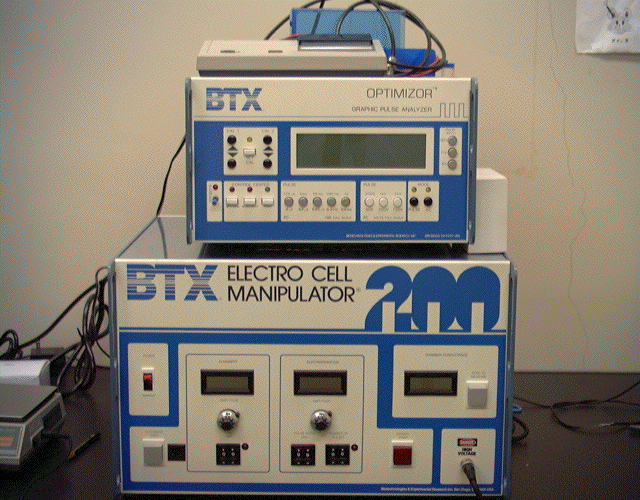
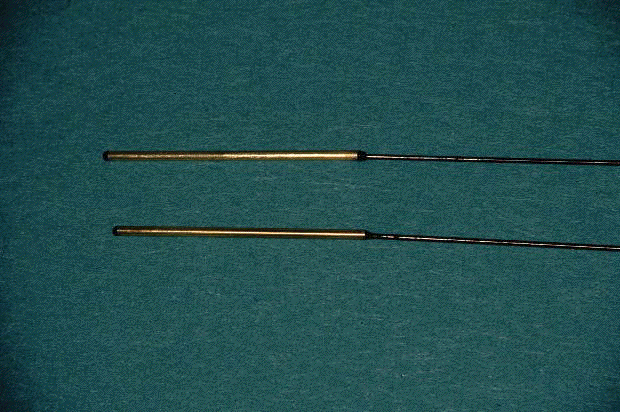
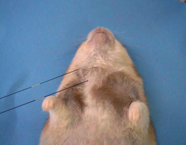
The electric pulses were derived from a square wave pulse generator (BTX electroporation unit, ECM 200, San Diego, CA, USA)(Fig. 1a) and applied at the tumor site using the stainless steel electrodes, 2 cm long and 0.5 mm in diameter, coated with gold (Fig. 1b) . The electric field was monitored during the treatment by a BTX Optimizer(BTX 500, San Diego, CA, USA) . The electrodes were inserted through the skin and placed at the bilateral margins of the metastatic lesion of the tumor(Fig. 1c). Tumor growth was followed by measuring three mutually orthogonal tumor diameters (a, b, and c) with vernier calipers on the day of the first treatment (volume of the tumor on the first day is designated as Vo) and every 2 days thereafter (volume of the tumor at N day is designated as Vn). Tumor volumes were calculated by the formula ą X a X b X c / 6, developed by Auerbach et al.(7). The tumor growth rate (T.G.R.) was calculated by means of the following formula, T.G.R.= Vn / Vo. The significance of the difference between the groups was determined by using Student's t-test. The tumors of all groups were examined histologically on 0, 2, 4, 8 , 10, 12 and 14th post-treatment day.
 Click to enlarge
Fig.1a: Photograph of BTX electroporation unit (ECM 500, USA)
 Click to enlarge
Fig.1b: Photograph of the stainless steel electrodes, 2 cm long and 0.5 mm in diameter, coated with gold.
 Click to enlarge
Fig.1c: Treatment view of the electrochemotherapy. The electrodes are inserted through the skin and placed at the bilateral margins of the metastatic lesion of the tumor.
| <= Introduction | MATERIALS & METHODS | Results => |
| Discussion Board | Next Page | Your Poster Session |