Invited Symposium: Stroke/Cerebral Vasospasm
| INABIS '98 Home Page | Your Session | Symposia & Poster Sessions | Plenary Sessions | Exhibitors' Foyer | Personal Itinerary | New Search |
Results
Contraction induced by hemolysate in rabbit basilar artery The initial significant contraction was obtained with 1% hemolysate (p < 0.05 vs. control) and further contraction was achieved by 10% hemolysate. We have shown in our previous study that increasing the level of erythrocyte lysate to 100% induced further contraction.(15) Thus, the maximum contraction was not obtained even at the highest concentration of hemolysate used. Therefore, in this study, only 0.01-10% hemolysate concentrations were tested.
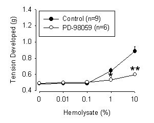
Effects of inhibitors on hemolysate-induced contractions Figure 1 shows the concentration-dependent contractions to hemolysate in the absence and presence of PD-98059. PD-98059 pre-incubated with rings for 30 minutes almost abolished the contractions to hemolysate (p < 0.05 -0.01, ANOVA). Since the maximum contraction to hemolysate was not obtained, we could not calculate the pA2 values. PD-98059 did not change the resting tension alone.
 click to enlarge
Fig. 1: Graphs displaying the inhibitory effects of the PD-98059. Arterial rings were contracted by exposure to hemolysate in the absence of PD-98059 in control rings. In treated group, PD-98059 (30 µM)was pre-incubated in separate chambers with arterial rings for 30 minutes before the rings were contracted with hemolysate. PD-98059 significantly inhibited contraction to 1% hemolysate (p = 0.018) (0.60 g vs. 0.53 g in control and 1% hemolysate, respectively) and to 10% of hemolysate (p = 0.002) (0.89 g vs. 0.60 g in control and 10% hemolysate, respectively) *p < 0.05, **p < 0.01 (ANOVA).
click to enlarge
Fig. 1: Graphs displaying the inhibitory effects of the PD-98059. Arterial rings were contracted by exposure to hemolysate in the absence of PD-98059 in control rings. In treated group, PD-98059 (30 µM)was pre-incubated in separate chambers with arterial rings for 30 minutes before the rings were contracted with hemolysate. PD-98059 significantly inhibited contraction to 1% hemolysate (p = 0.018) (0.60 g vs. 0.53 g in control and 1% hemolysate, respectively) and to 10% of hemolysate (p = 0.002) (0.89 g vs. 0.60 g in control and 10% hemolysate, respectively) *p < 0.05, **p < 0.01 (ANOVA).
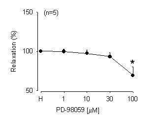
In another study, the rings of rabbit basilar artery were contracted with hemolysate (10%) and once a stable contraction was obtained, cumulative concentrations of PD-98059 was applied on the sustained contraction induced by hemolysate. PD-98059 produced significant relaxation (p < 0.05, ANOVA, Figure 2) at high concentration (100 µM).
 click to enlarge
Fig 2: Graphs displaying the relaxant effects of PD-98059 on sustained contraction induced by 10% hemolysate. When a stable contraction was obtained, PD-98059 was applied to relax the contracted arterial rings. PD-98059 significantly relaxed contracted artery in dose-dependent manner (p = 0.036). * p < 0.05 (ANOVA).
click to enlarge
Fig 2: Graphs displaying the relaxant effects of PD-98059 on sustained contraction induced by 10% hemolysate. When a stable contraction was obtained, PD-98059 was applied to relax the contracted arterial rings. PD-98059 significantly relaxed contracted artery in dose-dependent manner (p = 0.036). * p < 0.05 (ANOVA).
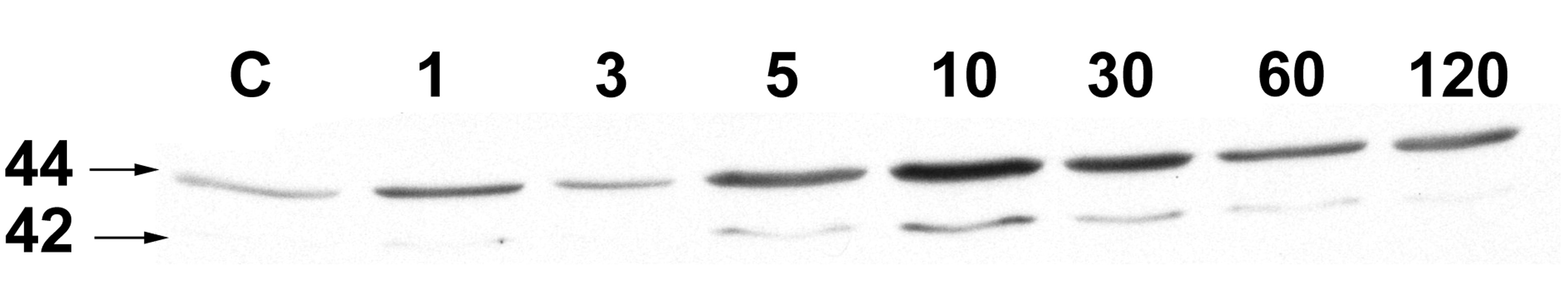
Hemolysate (10%) activates MAPK (ERK 1/2) in the first minute after exposure and induced peak MAPK enhancement at 5 minutes. The effect of hemolysate decayed with time but was still above the resting level even at 2 hours after exposure (Figure 3).
 click to enlarge
Fig 3: . Time course of activation of MAPK after stimulation with 10% hemolysate. A. MAPK was activated within 1 minutes, peaked by 5-10 minutes and maintained activity above baseline for at least 120 minutes. B. Graphs displaying the laser density of the protein bands showing quantified amount of activation of MAPK on different time point of stimulation. The peak of activation was at 5 minutes and activity of MAPK was maintained above baseline level for at least 120 minutes.
click to enlarge
Fig 3: . Time course of activation of MAPK after stimulation with 10% hemolysate. A. MAPK was activated within 1 minutes, peaked by 5-10 minutes and maintained activity above baseline for at least 120 minutes. B. Graphs displaying the laser density of the protein bands showing quantified amount of activation of MAPK on different time point of stimulation. The peak of activation was at 5 minutes and activity of MAPK was maintained above baseline level for at least 120 minutes.
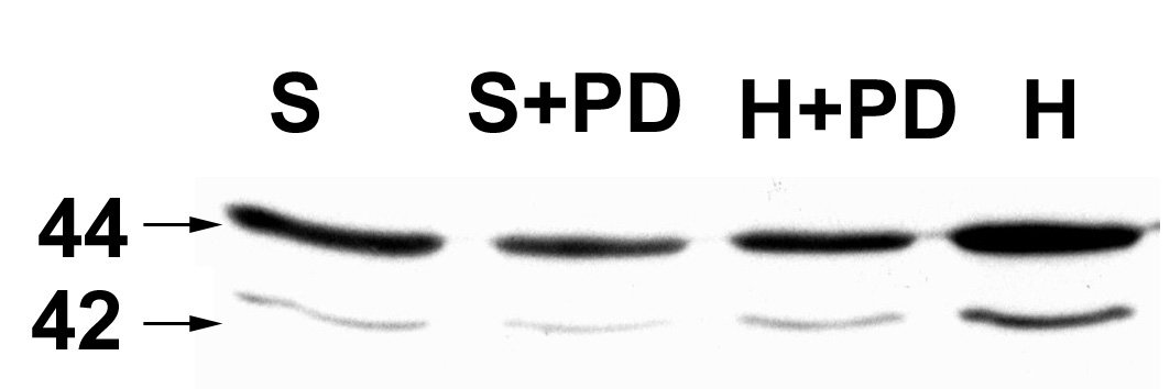
In a separate study, rabbit basilar arteries were treated for 30 minutes with saline or PD-98059 (30 µM) and then exposed to hemolysate (10%) for 5 minutes. Figure 4 demonstrates that hemolysate enhanced significantly MAPK phosphorylation and PD-98059 abolished the effect of hemolysate. PD-98059 did not markedly reduce the resting level of MAPK in rabbit basilar arteries.
 click to enlarge
Fig 4: A. Western blot picture demonstrating effect of PD-98059 on activation of MAPK after stimulation with hemolysate (10%). Incubation of control vessel with PD-98059 insignificantly decreased level of basal activity of MAPK. The effect of hemolysate (10%) on MAPK was abolished after the pre-incubation with PD-98059 for 30 minutes. B. Graph displaying the laser density of the protein band after the incubation with PD-98059. Non-significant decrease of basal level of MAPK activity appeared after the pre-incubation with PD-98059. Pre-incubation with PD-98059 for 30 minutes abolished the effect of hemolysate on MAPK (p = 0.041).
S - saline treated group of arteries, S+PD - vessels were pre-incubated with PD-98059 for 30 minutes and, then, treated with saline for 5 minutes. H+PD - vessels were pre-incubated with PD-98059 for 30 minutes and, then, treated with hemolysate (10%) for 5 minutes. H - vessels treated with hemolysate (10%) for 5 minutes.
click to enlarge
Fig 4: A. Western blot picture demonstrating effect of PD-98059 on activation of MAPK after stimulation with hemolysate (10%). Incubation of control vessel with PD-98059 insignificantly decreased level of basal activity of MAPK. The effect of hemolysate (10%) on MAPK was abolished after the pre-incubation with PD-98059 for 30 minutes. B. Graph displaying the laser density of the protein band after the incubation with PD-98059. Non-significant decrease of basal level of MAPK activity appeared after the pre-incubation with PD-98059. Pre-incubation with PD-98059 for 30 minutes abolished the effect of hemolysate on MAPK (p = 0.041).
S - saline treated group of arteries, S+PD - vessels were pre-incubated with PD-98059 for 30 minutes and, then, treated with saline for 5 minutes. H+PD - vessels were pre-incubated with PD-98059 for 30 minutes and, then, treated with hemolysate (10%) for 5 minutes. H - vessels treated with hemolysate (10%) for 5 minutes.
| <= Materials & Methods | RESULTS | Discussion & Conclussions => |
| Discussion Board | Next Page | Your Symposium |