Invited Symposium: Iron Transport
| INABIS '98 Home Page | Your Session | Symposia & Poster Sessions | Plenary Sessions | Exhibitors' Foyer | Personal Itinerary | New Search |
Results
Cellular survival and proliferation DTPA was the most effective Fe chelator at inhibiting cellular proliferation in all cell lines, with an IC50 value of 50ÁM in HTC cells (Table 1). This is in contrast to EDTA, another non permeable chelator from the same class which did not have any effect on cellular proliferation over the concentration range used (IC50 > 500ÁM). All three membrane permeable ferric chelators inhibited cell proliferation with L1 being the most effective (IC50 90ÁM), followed by DFO (IC50 180ÁM), then PIH (IC50 215ÁM). Of the ferrous chelators, the membrane permeable compound DP was active, inhibiting cell proliferation (IC50 160ÁM), whereas the membrane impermeable ferrous chelator BPS had little effect on cell proliferation (IC50 > 500ÁM).
Table 1: Effect of Fe chelators on proliferation of hepatoma cell line HTC
IC50 (ÁM)* CHELATOR DFO 180 PIH 215 L1 90 DTPA 50 EDTA >500 DP 160 BPS >500*[chelator] producing 50% inhibition
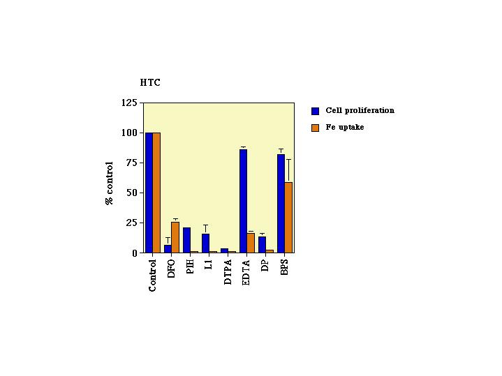
Effects of Fe chelators on Fe uptake: comparison with inhibition of cell proliferation Kinetic studies were performed to determine whether there was a correlation between the effects of the Fe chelators on cell proliferation and on Fe uptake into these cells. The level of inhibition of Fe uptake varied with cell line, incubation time, and chelator concentration. Representative data is shown for the HTC cell line over 24h at 500ÁM (Fig 1). For some chelators (eg BPS, DTPA) the degree of inhibition of cell proliferation was parallel to that of Fe uptake, but for other chelators (eg EDTA) there was no correlation
 Click to enlarge
Fig 1: Effect of chelators on Fe uptake and cell proliferation by the hepatoma cell line HTC. Cells were incubated with the chelator (500ÁM) for either 48hr (MTT assay) or 24hr (Fe uptake).
As illustrated in Fig. 1 Fe uptake and cell proliferation were both markedly reduced in the presence of the membrane permeable ferric chelators, PIH, L1 (and DFO). For the chelators PIH and L1, inhibition of Fe uptake was greater than cell proliferation. In contrast DFO had a greater effect on cell proliferation than Fe uptake. Interestingly, the impermeable chelator DTPA markedly reduced both Fe internalisation and cell proliferation. In contrast, the DTPA analogue EDTA had little effect on cell proliferation despite reducing Fe uptake by 80%. Of the ferrous chelators, the lipid permeable DP also greatly decreased Fe uptake, comparable to the level of inhibition of cell proliferation. In contrast, the membrane impermeable BPS had little effect on Fe uptake consistent with the low level of inhibition of cellular growth.
Effects of Fe chelators on Fe uptake; comparison with Fe uptake by normal hepatocytes
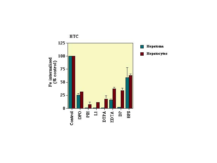
To determine whether there was any selectivity for neoplastic cells in the activity of the chelators, their effect on Fe uptake by hepatoma and hepatocyte cells were compared. In these experiments cultures of hepatocytes were also incubated for 24 hours with Tf-59Fe (1.25ÁM) in the presence or absence of the chelator (500ÁM).
 Click to enlarge
Fig. 2: Comparison of inhibition of Fe uptake by HTC cells and normal hepatocytes. Hepatoma cells (in exponential phase of growth) or hepatocytes were incubated with the chelator (500ÁM) for 24h.
The chelators PIH, L1, DTPA and DP were more active in HTC cells than in the hepatocytes, (Fig 2). However marked inhibition was also seen in the hepatocytes. The inhibition of Fe uptake by DFO was similar in the hepatoma cells and hepatocytes. EDTA was more effective at decreasing Fe uptake in hepatoma cells than hepatocytes and BPS had the least effect on Fe uptake in either cell type.
| <= Materials & Methods | RESULTS | Discussion & Conclussions => |
| Discussion Board | Next Page | Your Symposium |