Invited Symposium: Behaviour-Induced Neural Events after Brain Injury
| INABIS '98 Home Page | Your Session | Symposia & Poster Sessions | Plenary Sessions | Exhibitors' Foyer | Personal Itinerary | New Search |
Introduction
Previous studies have shown that following injury to the hand representation in primary motor cortex (M1), the size of the spared hand representation decreased substantially if monkeys recovered spontaneously (Nudo and Milliken, 1996). However, if monkeys received daily rehabilitative training on a task requiring skilled use of the impaired hand, the hand area spared by the lesion was retained (Nudo et al., 1996). To encourage these monkeys to use the impaired arm during rehabilitative training, the unimpaired arm was restrained.
The goal of the present study was to determine if restraint of the unimpaired forelimb in the absence of specific rehabilitative training was sufficient to retain spared hand area. The present study is important because it compares rehabilitation techniques similar to those used to treat human stroke patients. Studies involving stroke patients suggest that rehabilitative training of the impaired arm (physical therapy) combined with restraint of the unimpaired arm enhances long-term motor ability more than restraint of unimpaired arm alone (Taub and Wolf, 1997).
Materials and Methods
Three adult squirrel monkeys were employed in the present study. In each monkey, a map of the M1 hand area contralateral to the dominant hand was derived using standard intracortical microstimulation techniques. Then, a small (3-5 mm2) lesion was targeted to an area of primarily digit representations within the caudal portion of M1 using a bipolar coagulator, permanently sealing off blood supply to the target area. This destroyed ~30% of the M1 hand area (Fig. 1).
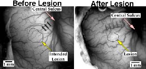 Fig. 1: Photographs of cortical surface before and one month after lesion. Shrinkage of the lesion area can be seen.
Immediately after the surgical procedure, a jacket with a long mesh sleeve over the unimpaired arm hand was placed on the monkey. Monkeys were required to retrieve small food pellets from wells of various sizes using their dominant hand (Fig. 2). The sleeve was sewn closed at the distal end. This sleeve allowed the animal to use proximal musculature (elbow, shoulder) freely but restricted use of the hand. The monkey was returned to its cage and was given no further intervention.
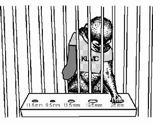 Fig. 2: Monkey with restrictive jacket performing pellet retrieval task (JACKET/REHABILITATIVE TRAINING group).
One month after the lesion, a second map of the M1 hand area contralateral to the dominant (impaired) hand was derived to assess changes in M1 topography following the month of hand restriction. In one monkey, additional maps were derived at three and six months after the lesion. The results from these three animals were compared to two other groups of animals from previous studies:
JACKET/NO TRAINING group (present results): underwent M1 mapping, lesion, and remapping sequence as outlined above. After lesion, wore restrictive jacket.
SPONTANEOUS RECOVERY group: underwent M1 mapping, lesion, and remapping sequence as outlined above for the JACKET/NO TRAINING group, but did not wear a jacket after the lesion (Nudo and Milliken, 1996).
JACKET/REHABILITATIVE TRAINING group: underwent M1 mapping, lesion, and remapping sequence as outlined above for the JACKET/NO TRAINING group, and wore a jacket after the lesion identical to those worn by the JACKET/NO TRAINING group. In addition, these animals received daily training on a pellet retrieval task for one month after the lesion (Nudo et al., 1996).
Results
One month after the lesion, the size of the total distal forelimb representation and the sizes of finger and wrist/forearm representations had decreased. Areal changes were significantly lower than in animals in a previous study that had received daily repetitive training after infarct (p<0.05). Areal changes were not different from animals that did not receive rehabilitative intervention or hand restraint after cortical injury (Figs. 3-5).
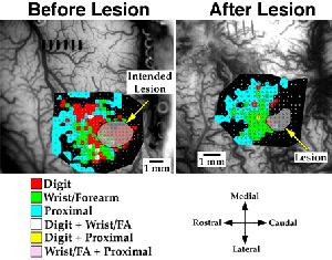 Fig. 3: ICMS-derived movement maps of M1 before and after lesion—SPONTANEOUS RECOVERY group. Proximal zones occupy portions of the former hand area.
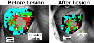 Fig. 4: ICMS-derived movement maps of M1 before and after lesion—JACKET/NO TRAINING group. Proximal zones occupy portions of the former hand area.
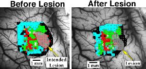 Fig. 5: ICMS-derived movement maps of M1 before and after lesion—JACKET/REHABILITATIVE TRAINING group. Digit and wrist areas outside the lesion are retained.
The spared digit, wrist/forearm, and total distal forelimb representational areas each decreased substantially in the JACKET/NO TRAINING group (Fig. 6). There were no statistical differences between areal changes of the JACKET/NO TRAINING (n=3) and SPONTANEOUS RECOVERY (n=3) groups. The JACKET/REHABILITATIVE TRAINING (n=4) group retained significantly more representational area than the other two groups. ANOVA p-values are shown in red for each group, significant Fisher’s post-hoc p-values are shown in blue. Error bars represent standard error of the mean.
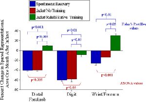 Fig. 6: Changes in spared representational areas one month after lesion.
Postlesion behavior is highly variable. It seems, however, that animals in the JACKET/REHABILITATIVE TRAINING group return to baseline efficiency on the task more quickly than animals in the other groups (Fig. 7).
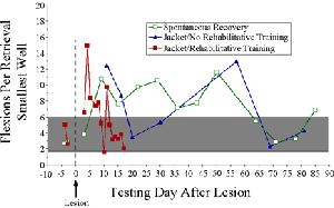 Fig. 7: Motor performance on the smallest well of the Klüver board after lesion.
Distal forelimb and digit areas increase gradually after lesion, suggesting that monkeys in the JACKET/NO TRAINING group may recover more slowly than monkeys in the JACKET/REHABILITATIVE TRAINING group (Fig. 8).
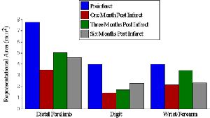 Fig. 8: Changes in representational area over time after lesion—JACKET/NO TRAINING group.
Discussion and Conclusion
Spared hand area was retained only in animals receiving repetitive training of the impaired hand. Monkeys whose unimpaired hand is restrained after cortical lesion may recover more slowly, both behaviorally and physiologically, than monkeys receiving rehabilitative training of the impaired hand. Thus, repetitive rehabilitative training of the impaired arm may be required for retention of undamaged hand area in M1. These results are similar to findings in human stroke patients-- rehabilitative training of the impaired arm combined with restraint of the unimpaired arm enhances long-term motor ability more than restraint of unimpaired arm alone (Taub and Wolf, 1997).
Acknowledgments:
We gratefully thank Archie Heddings, Erik Plautz, Scott Barbay, Ph.D., Shawn Frost, Ph.D., Cami Knox, Diane Larson, Haiying Wang, Gary Milliken, Ph.D., Frank SiFuentes, Birute Wise, Kathy Barbay, and Hector Sanchez for their assistance in completing this study. This work was supported by NIH NS 30853, AG 14635, and the American Heart Association (RJN).
References
- Nudo, R. J., & Milliken, G. W. (1996). Reorganization of movement representations in primary motor cortex following focal ischemic infarcts in adult squirrel monkeys. J. Neurophysiol., 75:2144-2149.
- Nudo, R. J., Wise, B. M., SiFuentes, F., & Milliken, G. W. (1996). Neural substrates for the effects of rehabilitative training on motor recovery after ischemic infarct. Science, 272:1791-1794.
- Taub, E., & Wolf, S. L. (1997). Constraint induced movement techniques to facilitate upper extremity use in stroke patients. Top. Stroke Rehabil., 3:38-61.
| Discussion Board | Previous Page | Your Symposium |