Poster
Contents
| INABIS '98 Home Page | Your Poster Session | Related Symposia & Posters | Plenary Sessions | Exhibitors' Foyer | Personal Itinerary | New Search |
Introduction
Increasing performance of digital image analysis and development of new classification algorithms, like neural networks, has led to new technological approaches in the diagnosis of skin cancer by analysing digitized images of clinical or dermatoscopic pictures. Taking into account epidemiological data and risk factors an automatic diagnostic system should allow to distinguish between benign and malignant pigmented skin lesions. To overcome the problem of a small amount of standardized data the in case of the DANAOS (Diagnostic And Neuronal Analysis of Skin Cancer) project, 14 European clinics are working together to collect standardized data of many thousands of suspicious skin lesions.Back to the top.
Camera System
Not only the amount but also the quality of the collected data is important to get a suitable training set for a neural network. Therefore a specially calibrated 3-Chip-Video-Camera (768x576 pixels) was constructed to aquire a set of standardized images in 10-,20- and 30-fold magnification. This high resolution allows the investigation of the fine texture of pigmented skin lesions. Several pictures can be shot in a short time interval under different lightening conditions by use of a ring of microprocessor controlled white-light-LEDs. These different illuminations can be used to analyse the the skin surface structure.
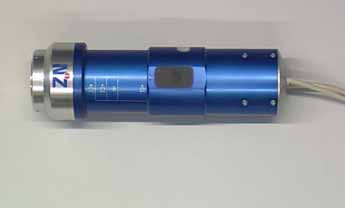 Fig. 1: Camera.
Database
A program was developed for rapid collection of consistant data of patient history and risk factors for malignant melanoma. The answers are controlled by several validation rules to minimize the risk of mistyped entries. The list of possible clinical and histological diagnosis was restricted to a few possible answers to give an easy way for training a neural network and for calculating sensitivity and specificity.
Fig.2: Inputmask for casehistory (ca. 86 kb)
click to enlarge
Fig.3: Inputmask for single lesion (ca. 97 kb)
click to enlarge
Results
On time we are testing several algorithms for extracting features according to the ABCDE-rule of dermatology. Especially the segmentation and the surface structure analysis have found to be of high accuracy.This is only filler text that is meant to fill out the poster and make sure that it takes up the full 80% of the page. This will ensure that it looks nice and will give a better idea of the finished state of the poster. There is liable to be quite a lot of text present in the real posters because there will be a lot of information to convey. There will be much coming to this spot in the future, and the sections will not all have the same amount of text.
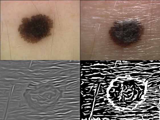
Fig. 5 a: ELM-picture (30x) b: sideward illumination
c:Surface structure d: Image c binarized. (ca. 180 kb)
Discussion
The camera sytem and the data aquisition unit was already delivered to 14 European dermatological departments. In this clinical units images and patient data were continuously collected and submitted to the Centrum for Neuroinformatic in Bochum/Germany. The developers in this centre are testing and improving the algorithms and training a neural network. We will present the results of this diagnostic unit at the end of this millenium.
| Discussion Board | Previous Page | Your Poster Session |