New Technology Poster Session
| INABIS '98 Home Page | Your Session | Symposia & Poster Sessions | Plenary Sessions | Exhibitors' Foyer | Personal Itinerary | New Search |
Introduction
To visualize the epidermal effects of different locally applied ointments and creams using sonography, an ultrasound transducer with a significant higher axial and lateral resolution than in the commercially available 20 MHz machines is necessary.
We used a 100 MHz transducer for this study.
This transducer has an axial resolution of 9 µm and a lateral resolution of 27 µm.
Since the stratum corneum is thicker than 100 µm on the finger pads, this transducer is well suited to study this particular skin area.
Materials and Methods
100 MHz-Sonography
The 100 MHz ultrasound imaging unit consists of an applicator with the transducer (PZT), a transient recorder, a control unit and a computer.
To account for the short depth of the focal area of the 100 MHz transducer a suitable imaging conception, the B/D(Brightness/Depth)-Scan concept is used.
B/D-Scan
A couple of "short" B-Scans is acquired at different depths of the tissue by mechanical movement of the transducer with respect to the depth direction (D-axis) between each acquisition.
Finally all scans are composed to get an image with a larger size. We call this additional axial motion "D-Scan" and thus, the whole procedure "B/D-Scan".
Applicator
Water is used as coupling medium. After placing the applicator on the skin, the plexiglas chamber is filled with water.
Three stepper motors are housed in the applicator which enable a precise movement of the transducer in all three axes.
Study design
We studied the effects of five different creams and ointments on the finger pads after occlusion of 10 healthy persons.
The occlusion was done with one square-centimeter Finn-Chambers.
The test sites were evaluated sonographically after an occlusion period of 0 (base line), 30, 60, 120, 180 and 240 minutes.
Creams and ointments
We chose the following creams and ointments:
vaseline, a water-in-oil-emulsion, an oil-in-water-emulsion and water.
They differ in their fat and water content.
When externals consist mainly of fat, the moisturization increases due to occlusion, i.e. the vaporisation is reduced.
The more water an external contains, the more swelling of the horny layer is seen due to direct water uptake.
10 % urea in an oil-in-water-emulsion was selected as 5th external.
Results
Figure 1 shows the sonogram of the finger pad before therapy:
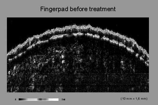
Figure 1
The top echorich line is the skin entry echo.
Then follows an upper echopoor band. This represents the stratum corneum.
A sweat gland duct in the stratum corneum is seen.
The lower echopoor band represents the stratum Malpighii and the stratum papillare.
Between the stratum corneum and the stratum Malpighii an echorich line is seen.
Beneath is the echoricher stratum reticulare of the dermis.
The thickening of the stratum corneum is already visible clinically.
Figure 2 shows the effect of the oil-in-water-emulsion on the same finger pad
after an occlusion of 30 minutes,

Figure 2
and figure 3 the corresponding sonogram after 30 minutes occlusion with the oil-in-water-emulsion.
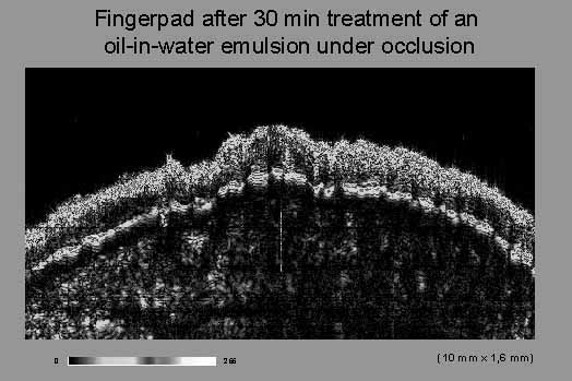
Figure 3
A significant thickness increase of the stratum corneum can be seen.
The echo density of the upper stratum corneum also increases.
Figure 4 summarizes the thickness increase of the stratum corneum at different time periods using the various ointments.
Figure 4: Thickness increase in µm after different occlusion periods: 0, 30, 60, 120, 180 and 240 minutes.
Externa: vaseline (red), water-in-oil-emulsion (green), oil-in-water-emulsion (dark blue),
10 % urea in the oil-in-water-emulsion (yellow), water (light blue).
The fastest and largest thickness increase of the stratum corneum is observed under the oil-in-water-emulsion and water.
After 240 minutes the thickness value for the oil-in-water-emulsion reaches 100 %.
When the external is more fatty (i.e. vaseline), swelling occurs later. But the end points are also quite remarkable.
Discussion and Conclusion
The thickness increase of the stratum corneum is due to an increase in humidity, which is a direct effect of water-containing externals or which is due to a reduction of vaporisation in fat-containing externals.
We observed the following effects of locally applied externals on the ultrasound image:
- a significant thickening of the stratum corneum on the finger pads,
- a change of the echo intensity of the upper stratum corneum and
- a reduction of the echogenicity of the skin entry echo after a long application time.
We conclude, that 100 MHz sonography therefore is well suited to evaluate the epidermal effects of topically applied substances in follow-up studies.
| Discussion Board | Previous Page | Your Poster Session |