Poster Contents
| INABIS '98 Home Page | Your Session | Symposia & Poster Sessions | Plenary Sessions | Exhibitors' Foyer | Personal Itinerary | New Search |
Results
There was no difference between old animals in the two species. The young animals (3-year old) showed no iron deposits. In the old animals (8-15-year old), Perl's staining without DAB intensification demonstrated the presence of numerous Prussian blue-positive cells in tissue sections (fig. 6). Iron pigments were mainly localized in the globus pallidus, the substantia nigra, the neocortical and cerebellar white matter, the thalamus, and the olfactory bulb. This distribution agrees with previous findings in humans (Hallgren and Sourander, 1958). Iron is also observed in basal forebrain structures including the nucleus accumbens, the septum, the diagonal band of Broca, the substantia innominata (which includes the nucleus basalis of Meynert) (Ch 1-4) and the olfactory tubercle.
Iron deposition in the aged mouse lemur basal forebrain has not been visualized at 4.7 T (Dhenain et al., 1997). At 11.7 T, the contrast is substantially enhanced and the effect of iron presence is very apparent (figs. 1-6).
The figures 1-5 show coronal MR scan T2*-weighted images of a 12-year old mouse lemur brain. The hypointense (dark) signal on the left images corresponds to iron accumulation and largely overlaps the distribution of ChAT-immunoreactive neurons. On the right images, the resulting views after thresholding display the septum, the diagonal band of Broca, the substantia innominata (figs. 1-3), and the globus pallidus (figs. 3-4). The substantia nigra is displayed when applying a slightly higher threshold (fig. 5). For the old animals (8-15 yrs old), the mean value of the upper threshold is 30.7 for the globus pallidus, 33 for the basal forebrain structures, and 41.3 for the substantia nigra.
These results indicate that the MR contrast resulting from the iron content is comparable in the basal forebrain structures and in the globus pallidus. Interestingly, the largest age-related increase in ferritin is seen in the medial septum of the rat (Focht et al., 1997). When applying the upper threshold at 35 or 4 1, midbrain structures, including the pedunculopontine tegmental cholinergic nucleus (Ch 5-6), are not displayed. It suggests that iron deposits affect basal forebrain cholinergic structures because of their close relation with the ventral globus pallidus. Further longitudinal studies are necessary to assess how correlated are the iron depositions in the basal forebrain and the globus pallidus.
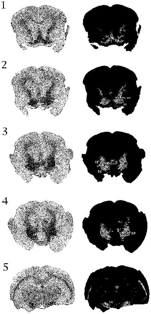 Fig. 1-5: Coronal MR scan T2*-weighted images of the 12-year old mouse lemur brain. The hypointense (dark) signal on the left images corresponds to iron accumulation in basal forebrain structures (bf) such as the septum, the diagonal band of Broca and the substantia innominata (figs. 1-3), in globus pallidus (gp) (figs. 3-4), and in substantia nigra (sn) (fig. 5). Iron is also present in anterior commissure (ac) and internal capsule (ic). Signal intensity was measured using a gray scale ranging from 0 to 255. On the right images, the voxel values beyond a defined upper threshold are eliminated. The upper threshold was set at 35 (figs. 1-4) and at 41 (fig. 5). The right images indicate that the contrast resulting from the iron content is almost similar in the basal forebrain structures and in the globus pallidus.
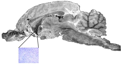 Fig. 6: Parasagittal MR scan T2*-weighted image of the 12-year old mouse lemur brain showing iron accumulation in the basal forebrain (dark signal) rostral to the anterior commissure (ac). This region comprises the septum, the nucleus accumbens, the diagonal band of Broca and the olfactory tubercle. The inset represents a high magnification of Perl's blue staining in a small portion of this region. Iron pigments (blue) are intracytoplasmic and surround the cell nuclei (pink). Scale bar = 25 microns.
| <= Materials & Methods | RESULTS | Discussion & Conclussions => |
| Discussion Board | Next Page | Your Poster Session |