Neuroscience Poster Session
| INABIS '98 Home Page | Your Session | Symposia & Poster Sessions | Plenary Sessions | Exhibitors' Foyer | Personal Itinerary | New Search |
Introduction
The two goals of this investigation were: 1) to determine if the transected distal stump of lumbar spinal nerves influences swelling of motoneuron cell bodies after axotomy; and 2) to characterize properties of the swelling response that can be used in studies of the mechanism of perikaryal enlargement after injury. The possibility that the severed distal stump might affect perikaryal swelling arose from reports that the distal stump is the source of several trophic factors (1-4) and cytokines (5,6) that increase in the vicinity of the axonal lesion. Thus, three properties of swelling - magnitude, laterality and uniformity - were quantified in the presence and absence of the distal stump.
Materials and Methods
The 9th and 10th spinal nerves of adult (60 ± 12 g) anesthetized grass frogs (R. pipiens) of both sexes were unilaterally transected about 5 mm distal to their exit from the spinal column. In those experiments involving stump removal, the severed nerves distal to the site of transection were dissected from the retroperitoneal space and thigh on the same side and removed. Seven days after axotomy, tissues were fixed by intracardiac perfusion and overnight immersion in cold 4% p-formaldehyde, and the control and experimental lumbar spinal cords were cryosectioned at 10µm and every fifth section was stained with cresylecht violet, and subjected to morphometric analysis. In each section examined, areas were defined for all motoneurons with a visible nucleolus, using a Technology Resources Imagemaster 1000 system calibrated with 9µm diameter plastic beads (Flow Cytometry Standards, Inc.). In the frog, lumbar motoneurons are easily distinguished by their size from other neurons in the ventral horn. Gamma motoneurons are not found in the frog spinal cord (7). The perimeter of each nucleolus, nucleus and cell body was traced three times with a mouse-driven cursor and the mean of the three computed areas was converted to µm2.
Results
I.The Swelling Response in Lumbar Motoneurons to Axotomy is Strictly Unilateral.
In the presence of the distal stump, the swelling response of motoneuronal nucleoli, nuclei and cell bodies is observed only on the operated side of the lumbar spinal cord. (Table 1). No significant size differences for these three structures were observed between contralateral and unoperated motoneurons.
Table 1. Nucleolar, nuclear and cell body areas (mean ± S.D.) in lumbar motoneurons 7 days after unilateral spinal nerve transection. Data from 7 operated frogs (331 motoneurons analyzed) and 6 unoperated frogs (446 motoneurons analyzed). a = p < 0.01 vs. contralateral values by Student's paired t-test (df = 6). b = p < 0.01 vs. unoperated controls by Student's unpaired t-test (df = 11).
                      Area(µm2), Distal Stump Left In
          Ipsilateral Contralateral Ipsi/Contra  Unoperated  Ipsi/Unop
Nucleolus   25.3± 3.4     15.9± 2.3     159.1%    17.6± 0.9     143.8%
              a,b
Nucleus    195.7± 36.3   183.6± 20.6    106.6%   173.3± 8.6     112.9%
               a
Cell body 1360.2± 192.8 1014.2± 94.7    134.1%  1040.5± 103.9   130.7%
              a,b
II. The Magnitude and Unilaterality of Swelling are Not Influenced by Removal of the Distal Stump.
Comparison of the data in Table 1 and Table 2 (below)shows that removal of the distal stump affects neither the magnitude of the mean swelling responses of nucleoli, nuclei or cell bodies on the ipsilateral side of the spinal cord nor the absence of a swelling response in contralateral motoneurons.
Table 2. Mean areas (± S.D.) in motoneurons 7 days following removal of distal stump (635 motoneurons from 15 frogs). a = p< 0.01 vs. contralateral values by Student's paired t-test (df = 6). b = p < 0.01 vs. unoperated controls by Student's unpaired t-test (df = 11).
                     Area(µm2), Distal Stump Removed
               Ipsilateral       Contralateral       Ipsi/Contra

Nucleolus        25.8± 2.1         16.9± 1.3             152.7%

Nucleus         215.9± 19.4       183.9± 17.7            117.4%

Cell Body      1453.9± 85.6      1098.6± 79.9            132.3%
III. Lumbar Motoneurons of All Sizes Swell Proportionately After Axotomy.
 click to enlarge
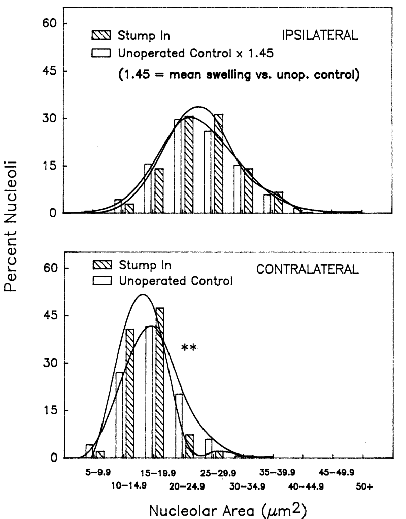
Fig. 1. Comparison of nucleolar area distributions in lumbar motoneurons of unoperated animals and axotomized animals in which the distal stump was present. The unoperated control values were obtained from both sides of the spinal cords of control animals. ** = p < 0.01 in chi-squared analysis.
click to enlarge
Fig. 1. Comparison of nucleolar area distributions in lumbar motoneurons of unoperated animals and axotomized animals in which the distal stump was present. The unoperated control values were obtained from both sides of the spinal cords of control animals. ** = p < 0.01 in chi-squared analysis.
 click to enlarge
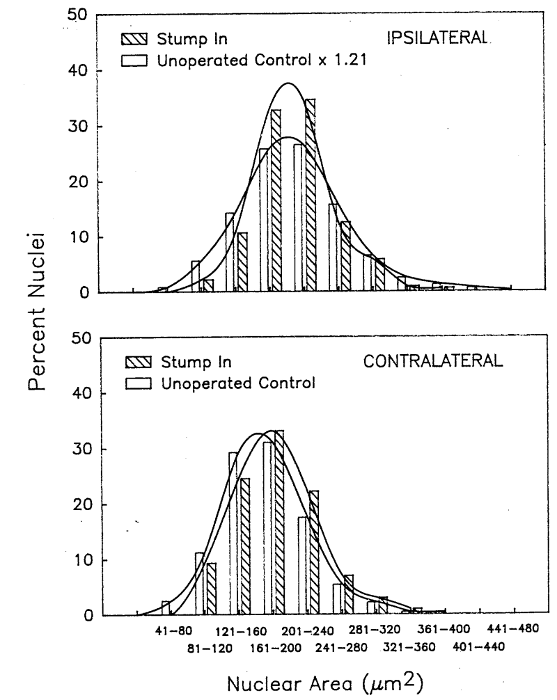
Fig. 2. Comparison of nuclear area distributions in lumbar motoneurons of unoperated animals and axotomized animals in which the distal stump was present. No statistically significant differences were found ipsilaterally or contralaterally in chi-squared analysis.
click to enlarge
Fig. 2. Comparison of nuclear area distributions in lumbar motoneurons of unoperated animals and axotomized animals in which the distal stump was present. No statistically significant differences were found ipsilaterally or contralaterally in chi-squared analysis.
 click to enlarge
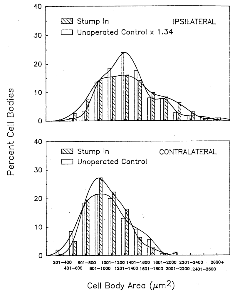
Fig. 3. Comparison of cell body area distributions in lumbar motoneurons of unoperated animals and axotomized animals in which the distal stump was present. No statistically significant differences were found ipsilaterally or contralaterally in chi-squared analysis.
click to enlarge
Fig. 3. Comparison of cell body area distributions in lumbar motoneurons of unoperated animals and axotomized animals in which the distal stump was present. No statistically significant differences were found ipsilaterally or contralaterally in chi-squared analysis.
In the upper panels of Figs. 1-3, the area of each individual unoperated control motoneuron was multiplied by the mean increase in area of the ipsilateral motoneurons. If spinal nerve transection caused every injured motoneuron to enlarge to precisely the same extent as the mean swelling of ipsilateral vs.unoperated neurons in Tables 1 and 2, then normalizing the control motoneuron data in this way would produce a frequency histogram of sizes that is superimposable upon the frequency histogram for the ipsilateral motoneurons. This is seen in Figs. 1-3 to be the case. Chi-squared analysis of the difference between the observed (stump in) and expected (unoperated control x factor) values indicated no significant difference in the distribution of nucleolar, nuclear or perikaryal sizes in injured and unoperated motoneurons.
When the size distributions of contralateral and unoperated motoneurons were compared (lower panels, Figs. 1-3), a significant difference (p < 0.01) was found by chi-squared analysis in the size distibutions of nucleoli in contralateral motoneuronal and unoperated controls (lower panel, Fig. 1), despite the absence of a significant difference between the mean values for those populations (Table 1). A significant difference was also found in chi-squared analyses for contralateral nucleoli from animals with the distal stump removed vs. unoperated controls (p < 0.01; data not shown). In each instance, there appeared to be a shift to the left in the frequency of contralateral nuclei with areas between 20-30µm2. It is thus possible that a slight shrinkage takes place in some motoneuron nucleoli contralateral to the site of injury under these experimental conditions. On the other hand, no differences were found in the size distributions of unoperated and contralateral nuclei or cell bodies (lower panels, Figs. 2 and 3).
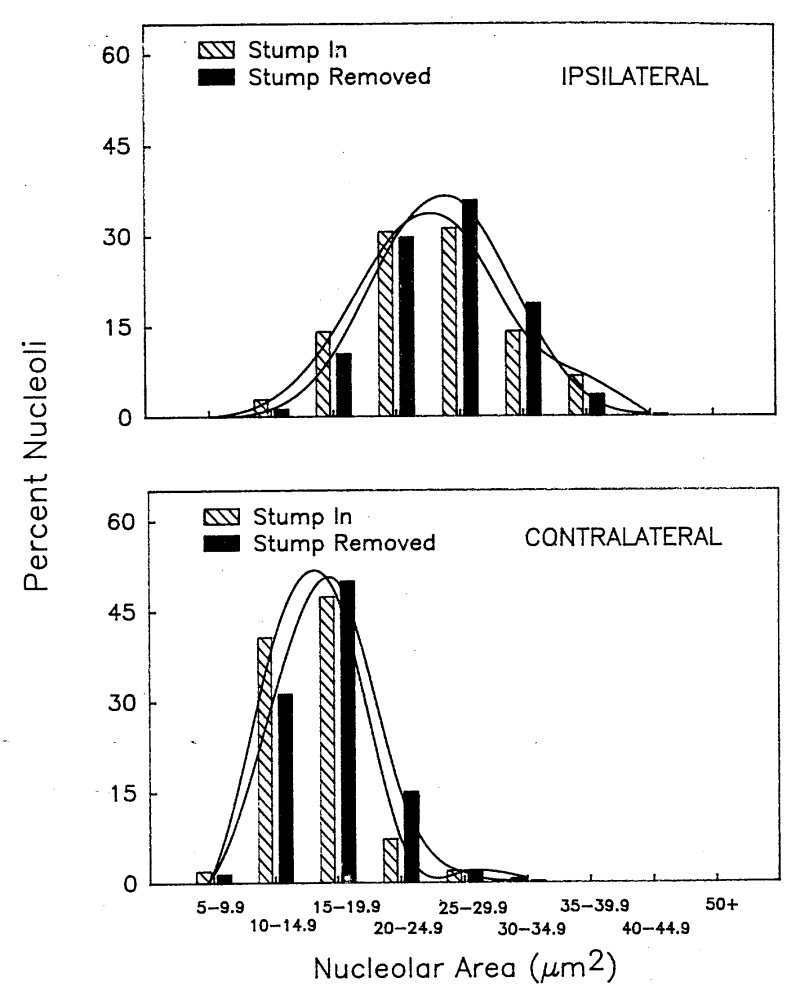
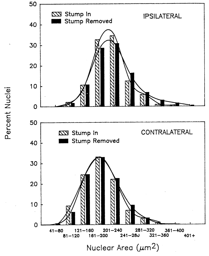
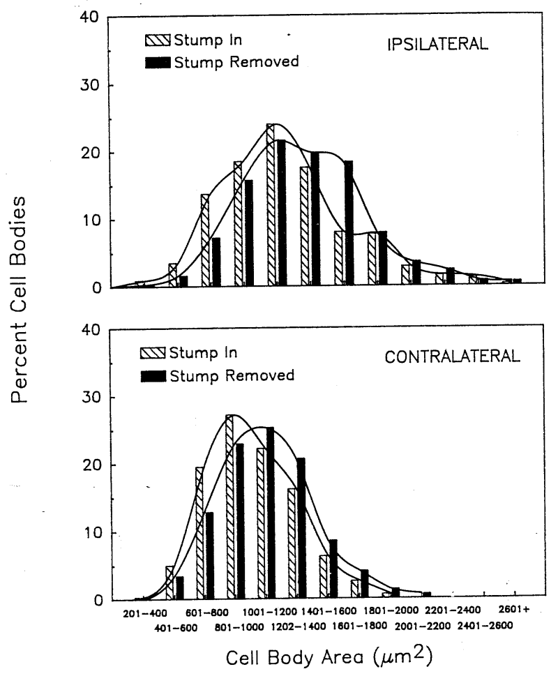
Figs. 4-6 show the results of similar analyses made after removal of the distal stump. No evidence was found by chi-squared analysis for differences in the size distributions of motoneuronal nucleoli, nuclei or cell bodies on either the ipsilateral or contralateral sides of the lumbar spinal cord when data from animals with and without distal stump removal were compared. Thus, both the mean areas and frequency histograms demonstrate that the magnitude and laterality of the swelling response to unilateral axotomy are not affected by removal of the distal stump.
 click to enlarge
Fig. 4. Comparison of nucleolar area distributions in ipsilateral and contralateral lumbar motoneurons in the presence and absence of the distal stump. No statistically significant differences were found ipsilaterally or contralaterally in chi-squared analysis.
click to enlarge
Fig. 4. Comparison of nucleolar area distributions in ipsilateral and contralateral lumbar motoneurons in the presence and absence of the distal stump. No statistically significant differences were found ipsilaterally or contralaterally in chi-squared analysis.
 click to enlarge
Fig. 5. Comparison of nuclear area distributions in ipsilateral and contralateral lumbar motoneurons in the presence and absence of the distal stump. No statistically significant differences were found ipsilaterally or contralaterally in chi-squared analysis.
click to enlarge
Fig. 5. Comparison of nuclear area distributions in ipsilateral and contralateral lumbar motoneurons in the presence and absence of the distal stump. No statistically significant differences were found ipsilaterally or contralaterally in chi-squared analysis.
 click to enlarge
Fig. 6. comparison of cell body area distributions in ipsialteral and contralateral lumbar motoneurons in the presence and absence of the distal stump. No statistically significant differences were found ipsilaterally or contralaterally in chi-squared analysis.
click to enlarge
Fig. 6. comparison of cell body area distributions in ipsialteral and contralateral lumbar motoneurons in the presence and absence of the distal stump. No statistically significant differences were found ipsilaterally or contralaterally in chi-squared analysis.
Discussion and Conclusion
The Role of the Distal Stump in Perikaryal Responses to Axotomy. The distal stump influences changes in axotomized motoneurons in a selective and still incompletely understood way. Wu et al. (8) found evidence that the distal stump prevents nitric oxide synthase induction in cervical motoneurons and promotes their survival after axotomy. Our data indicate that the distal stump does not initiate the swelling responses in injured lumbar motoneuron cell bodies. Moreover, a study by Ochs et al.(9) found that the distal stump does not initiate chromatolysis within the injured motoneuron cell bodies. Whether the distal stump is a source for other long-distance responses to axotomy, such as the increases in circulating cytokines and transcriptional responses in spared motoneurons, remains to be shown.
The Mechanism and Functional Significance of Swelling Responses in Axotomized Lumbar Motoneurons. Despite a century of study, most of the classic morphological responses of the neuronal cell body and dendrites to axonal injury have not been explained. Depending upon the specific neuron, those responses can include perikaryal, nuclear and nucleolar swelling or atrophy, chromatolysis, nuclear eccentricity and dendritic atrophy (10). With regard to the swelling phenomena described here, it is not known how they are produced and whether they support or interfere with regeneration of the injured motor axon. Our study indicates that, once identified, the mechanism for swelling under the present conditions will be refractory to removal of the distal stump. Furthermore, it will exist only ipsilaterally, will operate uniformly upon injured motoneurons of all sizes, and will produce coordinated changes in nucleolar, nuclear and perikaryal size consistent with the magnitude and time course of swelling observed here. Once understood, the swelling mechanism may then shed light on the relationship of this particular injury response to axon regeneration.
References
- Heumann, R., D. Lindholm, C. Bandtlow, M. Meyer, M.J. Radeke, T. Misko, E.M. Shooter, and H. Thoenen (1987) Differential regulation of mRNA encoding nerve growth factor and its receptor in rat sciatic nerve during development, degeneration and regeneration: role of macrophages. Proc. Natl. Acad. Sci. USA 84: 8735-8739.
- Sendtner, M., K.A. Stockli, and H. Thoenen (1992) Synthesis and localization of ciliary neurotrophic factor in the sciatic nerve of the adult rat after lesion and during regeneration. J. Cell Biol. 118: 139-148.
- Funakoshi, H., J. Frisén, G. Barbany, T. Timmusk, O. Zachrisson, V.M.K. Verge, and H. Persson (1993) Differential expression of mRNAs for neurotrophins and their receptors after axotomy of the sciatic nerve. J. Cell Biol. 123: 455-465.
- Gold, B.G. and P.S. Spencer (1993) Neurotrophic function in normal nerve and in peripheral neuropathies. In Neuroregeneration (A. Gorio, Ed.), pp. 101-122. Raven Press, New York.
- Jander S., J. Pohl, C. Gillen, and G. Stoll (1996) Differential expression of interleukin-10 mRNA in Wallerian degeneration and immune-mediated inflammation of the rat peripheral nervous system. J. Neurosci. Res. 43: 254-259.
- Gray, E.G. (1957) The spindle and extrafusal innervation of a frog muscle. Proc. R. Soc. Lond. B. Biol. Sci. 146: 416-430. Wu, W., K. Han, L. Li, and F.P. Schinco (1994) Implantation of PNS graft inhibits the induction of neuronal nitric oxide synthase and enhances the survival of spinal motoneurons following root avulsion. Exp. Neurol. 129: 335-339.
- Ochs, S., H. Booker, and W.E. DeMyer (1961) Note on the signal for chromatolysis after nerve interruption. Exp. Neurol. 3: 206-208.
- Lieberman, A.R. (1971) A review of the principal features of perikaryal responses to axon injury. Int. Rev. Neurobiol 14: 49-124.
- Lindholm, D., R. Heumann, M. Meyer, and H. Thoenen (1987) Interleukin 1 regulates synthesis of nerve growth factor in non-neuronal cells of rat sciatic nerve. Nature (Lond.) 330: 658-659.
| Discussion Board | Previous Page | Your Poster Session |