Poster
Contents
| INABIS '98 Home Page | Your Poster Session | Related Symposia & Posters | Scientific Program | Exhibitors' Foyer | Personal Itinerary | New Search |
| Title: Amebiano Hepatic
Abscess. Analysis morphologic and ultrastructural. Report of a case.
Contact Person: Alberto G Pizarro (rediegal@homonet.com.mx)
Clinical history: 36 year-old man with clinical report of diarrhea with snot and abdominal pain, three days he later present enlarged of liver of 10 cm, with jaundice+, pain in right hipochondrio, with pain to the tact, fever of 38.5 grades C., coluria+. Liver Rx identificated three cystic of 5 cm to 10 cm.,with irregular borders; two days he later died for liver failure and peritonitis. Autopsy: We identified three Amebic abscess in the liver: The patient had one great amebic abscess of 10 cm.,and two of 5 cm., Figure 1,figure 2; It contain yellow or gray, opaque liquid material. The wall was shaggy and fibrinous. Abscess has been complicated in hepatocolic fistula and perforation, causing peritonitis and death.
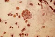
Microscopically.- The wall of abscess contained abundant fibrin. The trophozoites were abundant figure 3, figure 4 . The center was amorph,but it does not contain neutrophilic leukocytes. The surrounding liver was edematous.
Colonic amebic ulcers reavealed trophozoites on the surface of the mucosa, in the exudate, in the crater and submucosa. Figure 5 There is little inflammatory response of neutrophils and eosinophils.
|