Poster
Contents
| INABIS '98 Home Page | Your Poster Session | Related Symposia & Posters | Plenary Sessions | Exhibitors' Foyer | Personal Itinerary | New Search |
Introduction
The objective of this study was to quantify the UV transmission (Figure 1) of split skin exposed to UVB radiation and of non-exposed skin and to compare the two values.
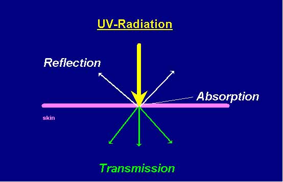
Figure 1
Materials and Methods
Two specimens of split skin having the dimensions 1 cm x 1 cm and with a thickness of 0.3 mm were taken from each patient.
In each case, one specimen was taken from an area of normal healthy skin and from an area that had been exposed to UVB radiation for 12 days. The initial dose of UVB was 1/3 MED (minimal erythema dose). The dose was raised by 1/3 MED every 4 days.
The figures 2 and 3 show the skin before and after 12 days of exposure to UVB-radiation.
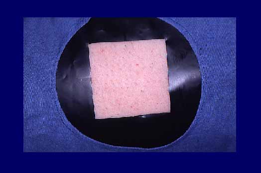
Figure 2
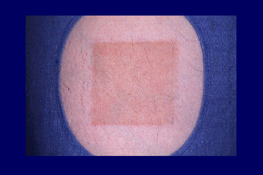
Figure 3
The split-skin specimens were streched until they exhibited the tension previously measured in vivo.
The UV transmission was measured with a Cary 3 Bio (VARIAN) spectrophotometer.
Results
Figure 4 shows the spectra of the mean transmission values obtained from the 12 patients for non-exposed skin and skin subjected to UVB radiation.
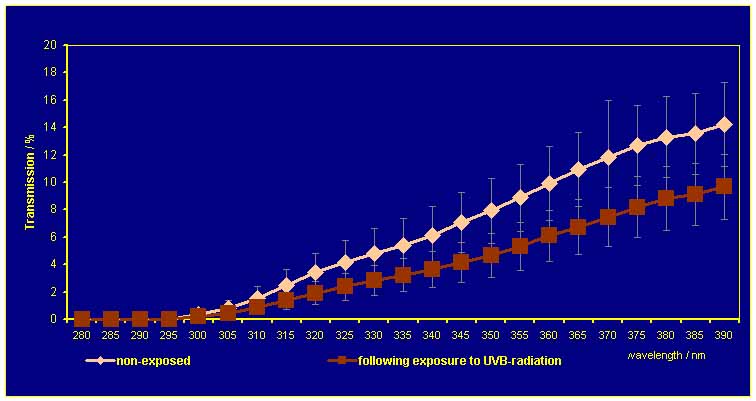
Figure 4
Figure 5 shows the mean transmission of non-exposed skin and split skin exposed to UVB radiation in the individual ranges: UVtotal (280-390 nm), UVA (315-390 nm) and UVB (280-315 nm).
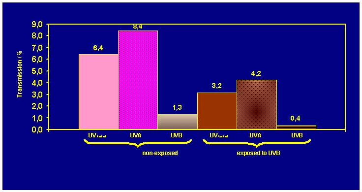
Figure 5
In the split-skin specimens exposed to UVB radiation, the mean transmission values were 50 % lower than in the non-exposed skin.
The ultraviolet protection factor (UPF) of the split- skin specimens was subsequently calculated from the transmission data.
The UPF of the split skin specimens of skin exposed to UVB radiation was found to be three times as high as the UPF of non-exposed skin (Figure 6).
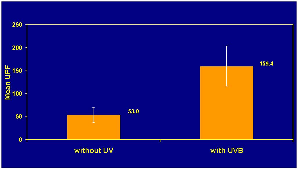
Figure 6
In figure 7 the results of the clinical determination of the minimal erythema dose (MED) in the course of the 12-day period of exposure to UVB (initial value, value after 6 days and value after 12 days) are shown.
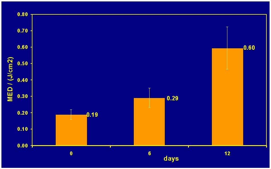
Figure 7
The MED of the patients was also raised by a factor of three by the 12 exposures to UVB-radiation. After only six exposures, the value rose by 1.5.
Discussion
The study had thus succeeded in objectifying and exactly quantifying the natural protection factor triggered by UVB radiation.
| Discussion Board | Previous Page | Your Poster Session |