Invited Symposium
| INABIS '98 Home Page | Your Session | Symposia & Poster Sessions | Plenary Sessions | Exhibitors' Foyer | Personal Itinerary | New Search |
Results
Blood Pressure
Compared to low salt treated Dahl-S rats, the high salt diet served to significantly elevate the mean blood pressure of Dahl-S rats (p < 0.0001), thus making these animals hypertensive. Mean blood pressures of high salt + tungsten treated Dahl-S rats were significantly lower than high salt treated Dahl-S rats (p < 0.05) but also significantly higher than low salt treated Dahl-S rats (p < 0.05). In contrast, mean blood pressures of low salt, high salt or high salt + tungsten treated Dahl-R rats did not differ (Table 1). Similar trends were seen in systolic and diastolic blood pressures.
Table 1
Central hemodynamic parameters of Dahl-R and Dahl-S rats after 4 weeks on either a low salt, high salt diet or high salt + tungsten diet.
---------------------------------------------------------------------
             Body Weight (g)   MBP (mm Hg)     SBP      DBP (mm Hg)
---------------------------------------------------------------------
Dahl-R
0.3% NaCl         327 ± 13      125 ± 5      157 ± 5     109 ± 5
6.0% NaCl         315 ± 19      133 ± 5      165 ± 5     117 ± 5
6.0% NaCl +       319 ± 19      133 ± 5      171 ± 12    115 ± 5
  0.07% Na2W04

Dahl-S
0.3% NaCl         359 ± 8       142 ± 8      174 ± 8     126 ± 8
6.0% NaCl         287 ± 42*     210 ± 15*    251 ± 20*   189 ± 15*
6.0% NaCl +       311 ± 43      176 ± 22*Ý   216 ± 17*Ý  156 ± 25*Ý
   0.07% Na2W04
---------------------------------------------------------------------
* p < 0.05 vs 0.3% NaCl group, Ý p < 0.05 vs 6.0% NaCl group.The mean arterial blood pressure in SHR under local anesthesia was significantly above normal as compared with that of WKY rats. This elevation in blood pressure was significantly lowered by the tungsten diet. In WKY rats, mean arterial blood pressure was in no case decreased but tended to show a marginal and insignificant increase after the tungsten diet. The systolic and diastolic blood pressures followed the same trend as the mean blood pressure (Table 2). There were no significant differences in heart rate among the four groups.
Table 2.
Systolic Blood Pressures (SBP), Diastolic Blood Pressures (DBP) and Heart Rate (HR) with and without Dietary Tungsten Supplement.
----------------------------------------------------------------------
                   WKY                           SHR
----------------------------------------------------------------------
            chow            tungsten        chow        tungsten
           (n=13)            (n=14)        (n=13)        (n=17)
----------------------------------------------------------------------
SBP     170.3 ± 26.6    185.1 ± 26.2    242.7 ± 45.3*   191.0±20.0#
(mmHg)
DBP     112.2 ± 11.6    121.9 ± 17.7    163.0 ± 10.4*   120.8±14.0#
(mmHg)
HR      409.3 ± 67.7    380.0 ± 46.3    396.6 ± 50.8   395.3 ±65.3
(bpm)
----------------------------------------------------------------------
Mean value ± SD.
n = number of animals.
*p<0.05 as compared with WKY rats (chow).
#p<0.05 as compared with SHR (chow).
Xanthine Oxidase
Xanthine oxidase activity in the Dahl strain rats was measured in spinotrapezius muscle samples since the mesentery was, unusable after the TNBT staining. Total skeletal muscle xanthine oxidase activity (XO+XD) in high salt + tungsten treated Dahl-S rats was significantly decreased compared to both low salt (p < 0.01) and high salt (p < 0.01) treated Dahl-S rats. Similarly, XO+XD activity in high salt + tungsten treated Dahl-R rats was significantly decreased compared to low salt (p < 0.01) and high salt treated Dahl-R rats (Table 3). In terms of xanthine oxidase activity (XO), again high salt + tungsten treated Dahl-S rats were lower than low salt (p < 0.05) and high salt (p < 0.01) treated Dahl-S rats and high salt + tungsten treated Dahl-R rats were lower than low salt (p < 0.05) and high salt treated Dahl-R rats (Table 3).
Table 3
Skeletal muscle xanthine oxidase activities of Dahl-R and Dahl-S rats after 4 weeks on either a low salt, high salt diet or high salt + tungsten diet.
----------------------------------------------------------------------
                         XO+XD(mU/mg)            XO(mU/mg)
----------------------------------------------------------------------
Dahl-R
0.3% NaCl                0.78 ± 0.07           0.33 ± 0.05
6.0% NaCl                0.73 ± 0.24           0.28 ± 0.13
6.0% NaCl                0.45 ± 0.10 *         0.16 ± 0.03 *
  + 0.07% Na2W04
Dahl-S
0.3% NaCl                0.91 ± 0.20            0.39 ± 0.20
6.0% NaCl                1.00 ± 0.23		0.35 ± 0.08
6.0% NaCl                0.54 ± 0.14 *#         0.18 ± 0.06 *#
  + 0.07% Na2W04
----------------------------------------------------------------------
* p < 0.05 vs 0.3% NaCl group, # p < 0.05 vs 6.0% NaCl group.XO and XD activity in the mesentery of SHR (1.94±0.77 mU/mg) was 2.8 times higher than that of WKY rats (0.70±0.15 mU/mg). The treatment with tungsten enriched diet lowered this activity to undetectable levels in both strains. XO activity in the mesentery of WKY rats (0.65±0.16 mU/mg) encompasses 93% of the total (XO and XD) activity (1.75±0.64 mU/mg); similarly, almost 90% of the total (XO and XD) activity in SHR is due to XO. The level of XO activity in the mesentery of SHR was 2.7 times higher than that of WKY rats. Treatment with a tungsten enriched diet reduced this activity in both strains to undetectable levels.
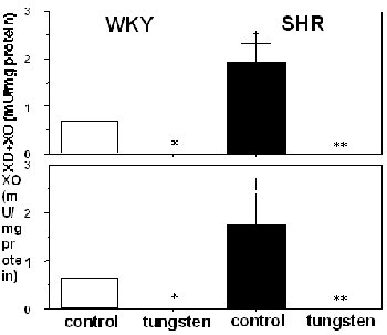 Figure 1. This figure shows the values of XO and XO+XD
In Vivo Superoxide Production
At a low magnification view of the mesentery preparation in the high salt Dahl-S rats we saw reduced TNBT, in the form of blue/black formazan crystals, along vessel walls of both arterioles and venules but not in the surrounding interstitial space. This is a feature we found uniformly in all hypertensive Dahl rats. Thin cross-sections of TNBT stained microvessels reveal dark formazan crystals within the vessel wall, not in the surrounding interstitial space and not inside the vessel itself, suggesting the endothelial cell is the actual site of superoxide generation.
Light micrographs of TNBT stained mesenteric microvessels from Dahl-R and Dahl-S rats revealed no significant difference in staining level between low salt, high salt and high salt + tungsten treated Dahl-R arterioles and venules. In contrast, an enhanced staining level was seen along the endothelium of arterioles and venules of high salt treated Dahl-S rats compared to low salt treated Dahl-S rats. The staining level in high salt + tungsten treated Dahl-S arterioles stained 20% lighter than high salt treated Dahl-S arterioles . In addition, high salt and high salt + tungsten treated Dahl-S venules stained 13% and 8% darker than low salt treated Dahl-S venules, respectively. By contrast, no significant difference in light absorption was observed between low salt, high salt and high salt + tungsten treated Dahl-R arterioles and venules.
(Dear Symposium Participant, we have tried to insert scanned images of the micrographs into this document. The image files are, however, too large and were not accepte d. Images are in the following references: Swei et al., Oxidative stress in the Dahl hypertensive rat. Hypertension, 30:1628-1633, 1997; Suzuki et al.; In-vivo evidence for microvascular oxidative stress in spontaneously hypertensive rats - Hydroethidine microfluorography. Hypertension, 25:1083-1089, 1995).
In the SHR, the level of HE oxidation, as demonstrated by the number of EB-positive nuclei (%) along mesenteric arteriolar walls, increased in SHR compared with WKY rats (Figure 2, left). The tungsten diet served to significantly decrease the number of EB-positive nuclei along arterioles of SHR (Figure 2, left), while in WKY rats, the tungsten diet had no effect on the number of EB-positive nuclei. In the venules, the level of HE oxidation was significantly higher in SHR than in WKY rats (Figure 2, right). The trend towards an elevated number of EB-positive nuclei along SHR venules was significantly attenuated after tungsten intake (Figure 2, right). In WKY rats, the tungsten diet had no significant effect on the number of EB-positive nuclei along the venules.
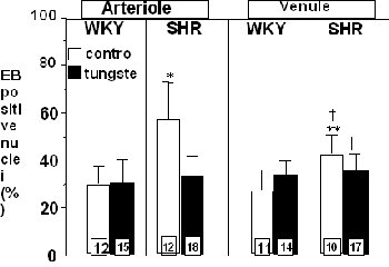 Figure 2. EB positve nuclei (%)
| <= Materials & Methods | RESULTS | Discussion & Conclussions => |
| Discussion Board | Next Page | Your Symposium |