Behavioural Neuroscience Poster Session
| INABIS '98 Home Page | Your Session | Symposia & Poster Sessions | Plenary Sessions | Exhibitors' Foyer | Personal Itinerary | New Search |
Introduction
Impairment of intellectual functions is a classic symptom of portal-systemic encephalopathy in humans. The hippocampal system is important for learning and memory both in humans and animals. Different subcortical neurotransmitter systems participate in the regulation of hippocampal activity and thus play a role in the cognitive process. It has been suggested that endogenous histamine, particularly neuronal histamine in the hippocampus and hypothalamus, may be involved in the process of learning and memory in rats.
Long-term portacaval anastomosis in the rat results in dramatic, region-selective changes in the brain histamine system, characterized by a marked increase in the tissue levels and a moderate elevation in the extracellular concentrations of histamine, and an increase in the tissue concentrations of tele-methylhistamine, most evident in the hypothalamus.
To examine the short-term and long-term effects of portacaval anastomosis on the hippocampal function in rats, we chose to study hippocampal EEG properties in rats with portacaval anastomosis at two consecutive time points, 1 month and 6 months after surgery. To disclose possible changes in the hippocampal histamine system, we assayed the tissue levels of histamine and tele-methylhistamine in the hippocampus of these rats 6 months after surgery.
Materials and Methods
Animal model. Male Wistar rats (initial weight 250 and 300 g) were anaesthetized with chloral hydrate (300 mg/kg; i.p.) and permanent electrodes were implanted in the hippocampus with the following coordinates relative to bregma: AP: -4.1; L: ± 2.6; V: -3.6. The electrodes were secured into the skull with dental acrylic cement and supported with four miniature stainless-steel screws fixed into the bone. PCA operation was performed according to the method of Lee and Fischer, 1961. Control rats were sham-operated using the same technique except for the anastomosis.
EEG recording and analysis. EEG from 17 sham-operated and 10 PCA-operated rats was recorded during the light phase, between 1 and 5 p.m, at two successive time points, 1 month and 6 months after surgery. EEG was sampled continuously for three hours at the rate of 120 Hz and the results were saved on the hard disk of the computer. The EEG data were analyzed off-line with the Matlab-Signal Processing Toolbox Software. Averaged power spectra were obtained with fast-Fourier transformations of EEG epochs, and the power densities of the 0-20 Hz frequency band and of the 5-9 Hz frequency band were computed.
Biochemical Assay. After completion of the experiment, 6 months after surgery, the rats were killed by decapitation and the hippocampi were dissected. Histamine in the hippocampus was assayed by HPLC coupled with fluorimetric detection; tele-methylhistamine by gas-chromatography-mass spectrometry.
Statistics. All data are expressed as means ± SEM. The Kruskal-Wallis test was used to measure the variability within the groups and to assess the significance of the group effects (PCA surgery and age). When two groups were compared, the Mann-Whitney U test was used. A probability of less than 5% (p<0.05) was considered to indicate statistical significance. Correlation analysis was performed by calculating the product-moment Spearman rank correlation coefficient.
Results
The results from the present study are presented in Figures 1 to 5.
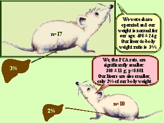 Figure 1. Mean body weight and liver-to-body weight ratio of sham-operated (n=17) and PCA-operated (n=10) rats six months after surgery. Statistics: Mann-Whitney U test.
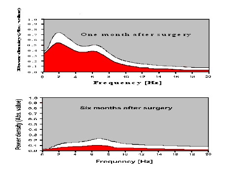 Figure 2. Averaged power density spectra of hippocampal EEG of sham-operated (n=17; white area) and PCA-operated (n=10; red area) rats 1 month and 6 months after surgery. Power density spectra were computed from the total 3 hours of EEG in each animal. Curve fittings were performed on the averaged spectra.
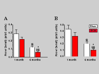 Figure 3. Power density of hippocampal EEG in sham-operated (n=17) and PCA-operated (n=10) rats, 1 month and 6 months after surgery. Values are expressed as arbitrary units and indicate mean ± SEM. Data were analyzed with the Kruskal-Wallis test, followed by the Mann-Whitney U test. * p<0.05 vs sham-operated controls; §§ p<0.01 vs. 1 month after surgery; §§§ p<0.001 vs 1 month after surgery. (A) Overall power density in the 0-20 Hz frequency band of hippocampal EEG. (B) Power density in the 5-9 Hz frequency band (theta rhythm) of hippocampal EEG.
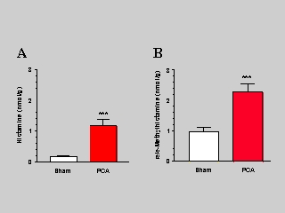 Figure 4. Concentrations (nmol/g wet tissue weight, mean ± SEM) of histamine (A) and tele-methylhistamine (B) in hippocampus of sham-operated (n=17) and PCA-operated (n=10) rats six months after surgery. Statistics: Mann-Whitney U test. *** p<0.001.
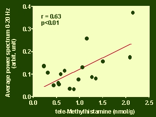 Figure 5. Scatter plot and correlation between the power density in the 0-20 Hz frequency band and the hippocampus tele-methylhistamine concentration of sham-operated rats 6 months after surgery. For each rat, the averaged power density was expressed in arbitrary units; histamine concentrations are given as nanomoles per gram of wet tissue weight. Statistical analysis was performed by calculating the product-moment Spearman rank coefficient (rs).
Discussion and Conclusion
PCA results in a severe liver atrophy and a significant reduction of the body weight of male Wistar rats.
Long-term PCA leads to a marked increase in the tissue levels of histamine and a moderate increase in the tissue levels of tele-methylhistamine in the hippocampus. These findings are suggestive of increased histaminergic activity in the hippocampus of rats with long-term PCA.
The frequency distribution of hippocampal EEG does not change significantly after PCA. However, the overall power density of hippocampal EEG and the power density in the 5-9 frequency band (theta rhythm) show a decreasing trend in the PCA rats, which has become significant 6 months after surgery. Since the electrophysiological properties have been regarded as important for the hippocampal function from the behavioural point of view, the reduced amplitude of hippocampal EEG in rats with portacaval anastomosis may represent the equivalent of impaired intellectual function in patients with portal-systemic encephalopathy.
A significant decrease in the amplitude of hippocampal EEG occurs with age. This decrease is particularly pronounced in the high-frequency range, including the 5-9 Hz frequency range, where the theta rhythm usually occurs. These time-related changes are more pronounced in the rats from the PCA group. It seems that, unlike the changes in neocortical EEG of PCA rats which once established remain, but do not progress with time, the EEG changes in the hippocampus of these rats are progressive.
Although inconclusive, the significant positive correlation between the levels of tele-methylhistamine, which reflect the amount of released histamine, and the power of hippocampal EEG in control rats, supports the notion that endogenous histamine may play a physiological role in modulating hippocampal EEG activity in rats. However, the present study does not provide evidence that the changes in the hippocampal histamine system contribute to the decrease in EEG activity induced by PCA in the rat.
| Discussion Board | Previous Page | Your Poster Session |