Invited Symposium: Behaviour-Induced Neural Events after Brain Injury
| INABIS '98 Home Page | Your Session | Symposia & Poster Sessions | Plenary Sessions | Exhibitors' Foyer | Personal Itinerary | New Search |
Results
Experiment 1
 Click to enlarge
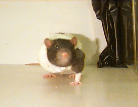
Fig. 1: Rat in limb-restricting cast designed to force reliance on one forelimb.
Click to enlarge
Fig. 1: Rat in limb-restricting cast designed to force reliance on one forelimb.
 Click to enlarge
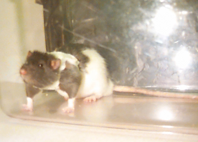
Fig. 2: Rat in a control cast designed to permit unrestricted movement of both forelimbs.
Click to enlarge
Fig. 2: Rat in a control cast designed to permit unrestricted movement of both forelimbs.
 Click to enlarge
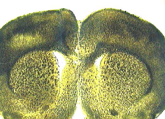
Fig. 3: Representative corpus callosum transection.
Click to enlarge
Fig. 3: Representative corpus callosum transection.
 Click to enlarge
Fig. 4: Diagram showing computer traced reconstruction of Layer V pyramidal neuron.
Total branch points, as well as branch points per branch order (see numbers on diagram) were counted.
Click to enlarge
Fig. 4: Diagram showing computer traced reconstruction of Layer V pyramidal neuron.
Total branch points, as well as branch points per branch order (see numbers on diagram) were counted.
 Click to enlarge
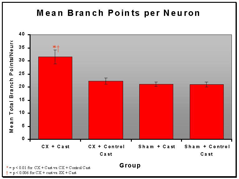
Fig. 5: Total branch points per neuron for each group. Animals with degeneration and behavioral demand (CX+Cast) had significantly higher branch points than animals with degeneration alone (CX + Control Cast; p<0.001) or behavioral demand alone (SX + Cast; p<0.006).
Click to enlarge
Fig. 5: Total branch points per neuron for each group. Animals with degeneration and behavioral demand (CX+Cast) had significantly higher branch points than animals with degeneration alone (CX + Control Cast; p<0.001) or behavioral demand alone (SX + Cast; p<0.006).
 Click to enlarge
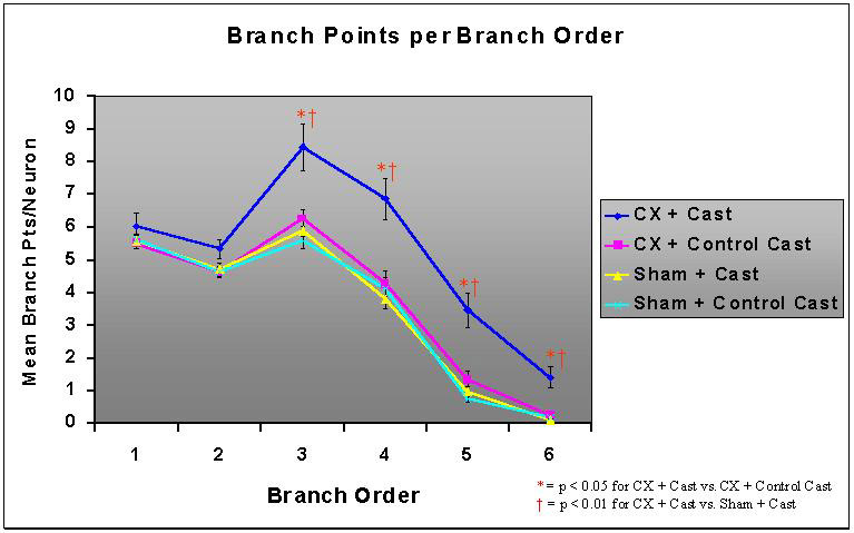
Fig. 6: Mean number of branch points per branch order. The increases in dendritic arborization in the CX + Cast group were greatest in higher order branches (third and higher).
Click to enlarge
Fig. 6: Mean number of branch points per branch order. The increases in dendritic arborization in the CX + Cast group were greatest in higher order branches (third and higher).
Experiment 2
 Click to enlarge
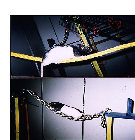
Fig. 7: Rats traversing two obstacles (a rope and a heavy link chain) of the acrobatic task. Rats were trained daily on the acrobatic task (adapted from Black, et al., 1990), which consisted of seven separate obstacles.
Click to enlarge
Fig. 7: Rats traversing two obstacles (a rope and a heavy link chain) of the acrobatic task. Rats were trained daily on the acrobatic task (adapted from Black, et al., 1990), which consisted of seven separate obstacles.
 Click to enlarge
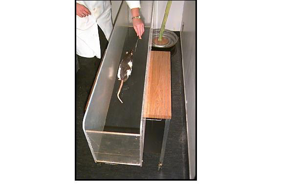
Fig. 8: Rats in the motor control group were given simple repetitive exercise on a straight runway.
Click to enlarge
Fig. 8: Rats in the motor control group were given simple repetitive exercise on a straight runway.
 Click to enlarge
Fig. 9: The sensorimotor cortex projects to the contralateral hemisphere of the cerebellar cortex via a multi-synaptic pathway. Thus, the hemisphere of the cerebellum contralateral to the lesion corresponds primarily to the impaired forelimb.
Click to enlarge
Fig. 9: The sensorimotor cortex projects to the contralateral hemisphere of the cerebellar cortex via a multi-synaptic pathway. Thus, the hemisphere of the cerebellum contralateral to the lesion corresponds primarily to the impaired forelimb.
 Click to enlarge
Fig. 10: Representative FLsmc lesion.
Click to enlarge
Fig. 10: Representative FLsmc lesion.
The Physical Disector Method: This technique was used to measure both synaptic density and Purkinje cell density. To determine density, adjacent sections were compared to identify synapses or cell bodies absent from one section that were present in the previous section. Tissue volume per neuron is the inverse of neuron density. Synapses per neuron is calculated as the product of synapse density and tissue volume per neuron.
 Click to enlarge
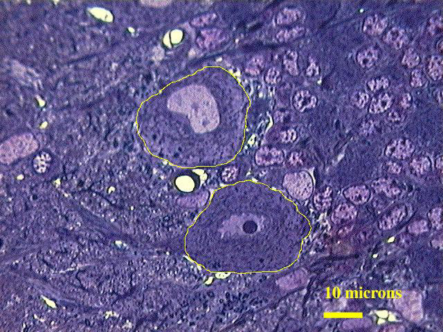
Fig. 11: The area of Purkinje cells was obtained by perimeter tracings of Purkinje cell bodies using Neurolucida software. Average Purkinje cell volume was calculated as the total volume fraction of cell bodies along the perimeter of the paramedian lobule (PML) divided by Purkinje cell density.
Click to enlarge
Fig. 11: The area of Purkinje cells was obtained by perimeter tracings of Purkinje cell bodies using Neurolucida software. Average Purkinje cell volume was calculated as the total volume fraction of cell bodies along the perimeter of the paramedian lobule (PML) divided by Purkinje cell density.
 Click to enlarge
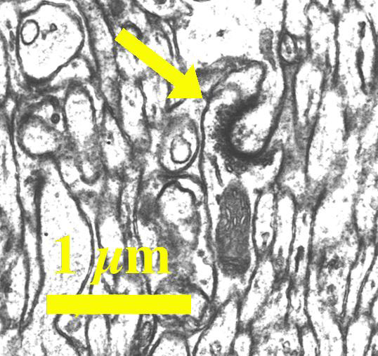
Fig. 12: Electron micrograph showing a synapse in the cerebellar molecular layer.
Click to enlarge
Fig. 12: Electron micrograph showing a synapse in the cerebellar molecular layer.
 Click to enlarge
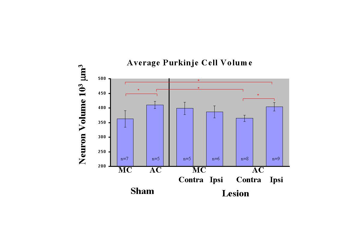
Fig. 13: Average Purkinje Cell Volume: Sham-operated acrobats (Sham-AC) had increased average Purkinje cell volume compared to the sham-operated motor controls (Sham-MC). In the lesion group, acrobats (Lesion AC) had increased cell volume in the PML ipsilateral to the lesion but not contralateral, relative to Sham-MC. The motor controls with lesions (Lesion-MC) showed a non-significant tendency toward increased Purkinje cell volume in both hemispheres.
Click to enlarge
Fig. 13: Average Purkinje Cell Volume: Sham-operated acrobats (Sham-AC) had increased average Purkinje cell volume compared to the sham-operated motor controls (Sham-MC). In the lesion group, acrobats (Lesion AC) had increased cell volume in the PML ipsilateral to the lesion but not contralateral, relative to Sham-MC. The motor controls with lesions (Lesion-MC) showed a non-significant tendency toward increased Purkinje cell volume in both hemispheres.
 Click to enlarge
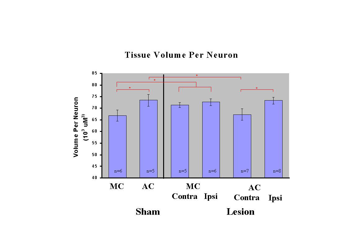
Fig. 14: Tissue Volume Per Neuron: Similar effects were found for tissue volume per neuron, with increased tissue volume (lower cell density) in Sham-AC relative to Sham-MC. In Lesion-AC rats, the ipsilateral but not contralateral PML was increased relative to Sham-MC. Lesion-MC showed elevations in tissue volume in both hemispheres, which was significantly greater than Sham-MC.
Click to enlarge
Fig. 14: Tissue Volume Per Neuron: Similar effects were found for tissue volume per neuron, with increased tissue volume (lower cell density) in Sham-AC relative to Sham-MC. In Lesion-AC rats, the ipsilateral but not contralateral PML was increased relative to Sham-MC. Lesion-MC showed elevations in tissue volume in both hemispheres, which was significantly greater than Sham-MC.
 Click to enlarge
Fig. 15: Synapses Per Neuron: No significant differences were found in synaptic density nor synapses per neuron. However, Sham-AC showed a tendency toward increased synapses per neuron consistent with previous studies (e.g. Black et al., 1990), while the Lesion-AC did not show this tendency.
Click to enlarge
Fig. 15: Synapses Per Neuron: No significant differences were found in synaptic density nor synapses per neuron. However, Sham-AC showed a tendency toward increased synapses per neuron consistent with previous studies (e.g. Black et al., 1990), while the Lesion-AC did not show this tendency.
| <= Materials & Methods | RESULTS | Discussion & Conclussions => |
| Discussion Board | Next Page | Your Symposium |