Poster
Contents
| INABIS '98 Home Page | Your Poster Session | Related Symposia & Posters | Plenary Sessions | Exhibitors' Foyer | Personal Itinerary | New Search |
Contact Person: Michel Ladouceur (mla@smi.auc.dk)
Materials & Methods
Participants
Eight participants (2 females, 6 males) took part in this study. All
had no diagnosed neurological or orthopaedic impairments. Their age ranged
from 25 to 32 years. All subjects gave their informed consent prior to
the experiments.
Experimental Set-up
Ankle stretches
The participants were seated in an adjustable chair with the left foot
firmly strapped to a foot plate. Knee and ankle joints were extended to
approximatively 1,5 rad. The ankle was rotated by a DC motor (CEM, model
26), connected to the foot plate. The axis of the rotation of the ankle
joint was aligned with the axis of rotation of the foot plate. The imposed
movements were a 0,07 rad dorsiflexion ramp followed by a subsequent hold
phase of 500 ms. The stretch velocity of the ankle extensors was varied
by changing the duration of the reference ramp signal. The ankle moment
was measured using strain gauges mounted on a beam connecting the foot
plate with the motor. The angular position of the foot plate was measured
by a potentiometer. The DC motor was powered by a DC amplifier (Bruel and
Kjær, Model 2708) and controlled by a position servo system. The
onset of the actual movement was delayed by 4 ms in respect to the reference
position signal supplied to the servo controller. Further details about
this setup have been described in elsewhere (Sinkjær et al., 1988).
Electromyography
The electromyogram (EMG) signals of the soleus (SOL) and tibialis anterior
(TA) muscles were recorded using bipolar surfce electrodes. A reference
electrode was placed above the knee. The EMG signals were amplified and
filtered (first-order band-pass filter: 20 Hz-1 kHz; DISA, model 15C01).
Digital processing of the data
Ankle angle and moment as well as amplified and filtered SOL and TA
signals were sampled at 2kHz and stored for further analysis.
The EMG signals were further processed by rectifying, low-pass filtering
(20 Hz) and averaging the signal after the end of the experimental condition.
In the analysis, only stretch reflexes acquired in random order in the
same test were compared. This reduced the effects of possible time dependencies
of the stretch reflex and conditioning processes.
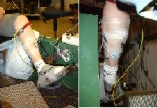
Fig. 1: The position of the EMG electrodes and the stretch device
Electrical stimulation of the nerves
The deep, superficial and common peroneal nerves as well as the sural
nerve were stimulated (Axon Instruments: Isolator 11 - stimulus isolation
unit) with a round 3 cm diameter self adhesive cathode placed on the skin
over the respective nerves. An oval anode electrode
(4*6 cm) was placed midway on the shank on the skin above the tibia.
The nerves were stimulated with trains of 5 pulses at 200 Hz, with a pulse
width of 1 ms. This stimulation frequency is markedly above the fusion
frequency of the TA, maximizing the activation of afferent nerve fibers,
while obtaining a fused TA contraction by the activation of motor fibers
in the deep and common peroneal nerves. The stimulation strength was controlled
by the stimulation current amplitude, which was manually adjustable on
the current controlled stimulator. The timing of the stimulation was controlled
by the PC that supplied the reference signal to the ankle stretching device
as well as digitising the different recorded signals.
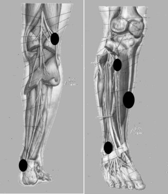
Fig. 2: Position of the conditioning electrodes
Experimental Conditions
The experiments were done over a two day period.
Comparaison of peroneals and sural nerve for the conditioning of
the SOL stretch reflex
On the first day, the stretch reflex inhibition in the SOL was tested
for the four different conditioning sites. For each stimulation site, the
interval between the conditioning stimulus burst and the onset of the triceps
surae stretch was varied. The tests were performed in a pre-contracted
triceps surae (5Nm). The deep and common peroneal nerves were stimulated
at 4,0 times TA motor threshold (MT) whereas the superficial peroneal and
sural nerve were stimulated at 4.0 times sensory threshold (ST). Stimulation
of up to 4 * MT for the deep and common peroneal nerve was justified by
the study of Gracies et al.(1994), who showed that additional recruitment
of Ia afferent nerve fibers can take place up to this stimulation level,
when using electrodes on the skin. For each stimulation site, the interval
between the beginning of the conditioning stimulus burst and the onset
of the stretch was varied between 50 ms and 1000 ms in 13 steps (50, 100,
150, 200, 250, 300, 400, 500, 600, 700, 800, 900, 1000 ms). Additionally,
stimulation without subsequent stretch and stretches without conditioning
stimulation were applied. All 15 conditions (13 delays, only stretch, only
stimulation) were applied approximately seven times in random order. A
20 ms stretch rise time was used for the delay tests, resulting in a maximal
stretch velocity of 2,025 rad/s.
Effect of four levels of triceps surae pre-contraction on the conditioning
from a common peroneal nerve stimulation
The inhibition of the stretch reflex at four different level of pre-contraction
(0, 2,5, 5, 10 Nm) was also tested on the first day. For this experimental
condition the common peroneal nerve was stimulated at 4.0 MT with a varying
conditioning-test interval (13 steps from 50 to 1000ms).
Effect of four intensities of conditioning stimulus on the stretch
reflex-velocity relationship
On the second day, the inhibition of the SOL stretch reflex was tested
for four different intensities of the conditioning stimulus (1,0, 2,0,
3,0, 4,0 MT) applied to the common peroneal nerve. The effect of the different
intensities were assessed at five different conditioning-test interval
(50, 100, 200, 300, 400 ms) as well as for six different stretch velocities
(0,438, 0,576, 0,846, 1,096, 1,536, 2,025 rad/s). These tests were performed
in a pre-contracted triceps surae muscle (5Nm).
Effect of a common peroneal nerve block on the conditioning of the
SOL stretch reflex
The inhibition of the SOL stretch reflex was also investigated after
a common peroneal nerve block. In one participant, the block of transmission
in the peroneal nerve was obtained by injecting lidocaine (10 ml; 0.5 mg/ml)
around the nerve at the level of the head of the fibula. Efficient block
of transmission was determined by the inability of the participant to voluntarily
activate the ankle dorsiflexors. Furthermore, the afferent inflow from
the stimulation was controlled by recording the sensory evoked potential
from the conditioning simulus.
Outcome measures
The outcome measure for this study was the peak of the SOL stretch
reflex and was determined from the rectified, filtered and averaged SOL
EMG signal. Furthermore, all the peaks were normalised to the unconditioned
values in order to compare the results across participants.
| <= Introduction | MATERIALS & METHODS | Results => |