Cell Biology Poster Session
| INABIS '98 Home Page | Your Session | Symposia & Poster Sessions | Plenary Sessions | Exhibitors' Foyer | Personal Itinerary | New Search |
Results
Immunohistochemistry
In the decidual stroma, interstitial trophoblasts were identified by the dense staining of the DAB reaction product [Figure 1a] indicating immunolabelling for cytokeratin. Immunolabelling for cytokeratin was also evident in the columnar epithelial cells of the endometrial glands but these cells could readily be identified by their distinct morphology. The cytokeratin labelling of epithelial cells served as an internal control.
Staining for neutrophil elastase was observed on numerous cells in regions of necrosis and around some of the maternal blood vessels [Figure 1b]. Some cells in the regions of necrosis were also positive for leucocyte common antigen. CD56 positive natural killer cells and HAM-56 positive macrophages were scattered around the decidual stroma and in close apposition with the interstitial trophoblast. The interstitial trophoblast cells were nonreactive with anti-CD56, anti-HAM-56 and anti-leucocyte common antigen. No significant number of neutrophils labelled with anti-neutrophil elastase was observed in areas of trophoblast invasion. No labelling was observed on negative control sections where the primary antibody was replaced by a non-immune serum.
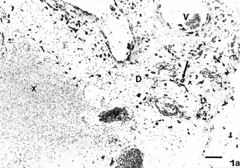 Fig. 1a: Photomicrograph of first trimester human decidua basalis immunolabelled with mouse monoclonal anti-cytokeratin. The reaction product of DAB is seen on the interstitial trophoblast cells (arrow) in the decidual stroma (D). The maternal leucocytes present in the region of necrosis are unreactive (X). Maternal blood vessels (V).
(Mag: x130, Bar: 100 um).
Fig. 1a: Photomicrograph of first trimester human decidua basalis immunolabelled with mouse monoclonal anti-cytokeratin. The reaction product of DAB is seen on the interstitial trophoblast cells (arrow) in the decidual stroma (D). The maternal leucocytes present in the region of necrosis are unreactive (X). Maternal blood vessels (V).
(Mag: x130, Bar: 100 um).
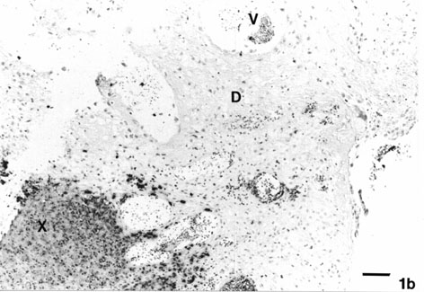 Fig. 1b: An adjacent section immunolabelled with anti-neutrophil elastase. The reaction product of New Fuchsin is confined to the neutrophils in the region of necrosis (X) and around some of the maternal blood vessels (V). Decidual stroma (D).
(Mag: x130, Bar: 100 um).
Fig. 1b: An adjacent section immunolabelled with anti-neutrophil elastase. The reaction product of New Fuchsin is confined to the neutrophils in the region of necrosis (X) and around some of the maternal blood vessels (V). Decidual stroma (D).
(Mag: x130, Bar: 100 um).
Ultrastructure
Thin sections from the basal decidua demonstrated two different interstitial trophoblast cell populations whose ultrastructure was distinct from those of decidual stromal cells and from decidual natural killer cells and maternal macrophages. The cells could be classified as, i) type I electron-lucent agranular [Figure 2a] and, ii) type II electron-dense granular interstitial trophoblast [Figures 2b and 2c]. The morphological features of the cells are summarised in table 1. Briefly, the type I cells represented a large proportion of the interstitial cell population compared to the type II cells. The former showed marked polymorphism, most cells appeared elongated and contained a centrally positioned nucleus with deep crenations and a prominent nucleolus. In areas where type II cells were abundant, apoptotic cells were also present. These were in the early stages of apoptosis or secondary necrosis [Figure 2b].
The cell membrane of the type I cells possessed numerous processes [Figure 3]. Their cytoplasm was electron dense containing numerous fine filaments, glycogen particles, well-developed stacks of Golgi apparatus, dilated rough endoplasmic reticulum and, numerous small mitochondria with a few cristae [Figures 3 and 4].
In contrast to the type I cells, the type II cells were near spherical in shape. Most cells had an eccentric nucleus with electron dense chromatin around the nuclear periphery. The most conspicuous feature of type II cells were the numerous membrane bound small electron dense granules scattered throughout the cytoplasm [Figures 2c]. The number of electron dense granules varied depending on the plane of section.
The ultrastructural morphology of the type II cells was closer to the morphology of the peripheral blood neutrophils than the type I cells. Nevertheless, unlike neutrophils, the type II cells were reactive for cytokeratin. Neutrophils were only present in the regions of necrosis and not in the decidual stroma.
The two interstitial trophoblast cell populations were present only in areas of basal decidua and not in the decidua parietalis (area away from the implantation site). In comparison to the type I cells, the type II cells contained a lobed nucleus. Their cytoplasm, unlike type I cells, contained less organelles and very few glycogen particles.
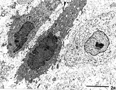 Fig. 2a: Electron micrograph of the first trimester human decidua basalis showing the two cell types. A decidual stromal cell (D) with a single nuclear profile, a prominent nucleolus and an electron lucent cytoplasm. The other two cells are type I agranular interstitial trophoblasts (T). They have an electron dense cytoplasm, a single large convoluted nuclear profile (n) and numerous cell processes (arrowhead). (Mag: x6410, Bar: 5 um).
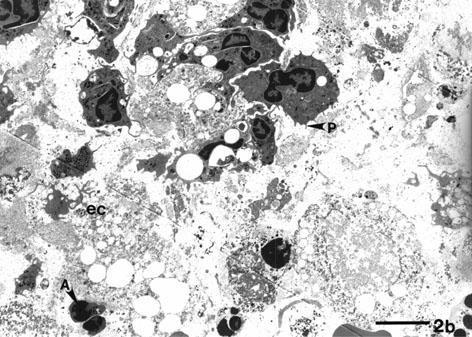 Fig. 2b: The type II granular interstitial trophoblast cells. The cells have numerous outer cell processes (P). There is extensive degenerative cellular material in the extracellular space (ec). There are also a few cells in this area with features suggestive of apoptosis (A) or secondary necrosis (S).(Mag: x1941, Bar: 10 um).
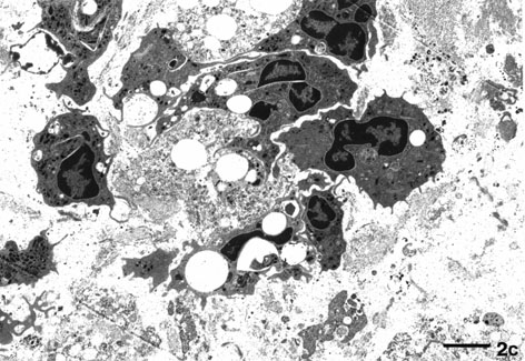 Fig.2c: A higher magnification showing several of the type II granular interstitial trophoblast cells. Their cytoplasm is electron dense and contains many granules.
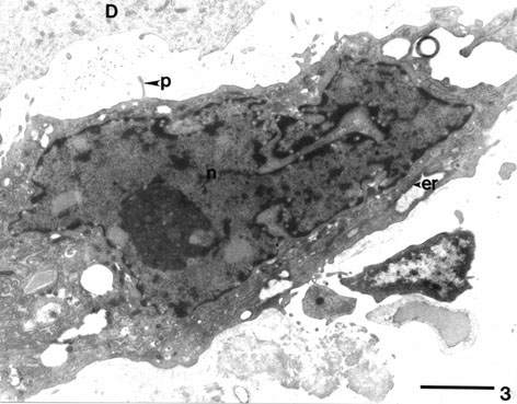 Fig. 3: An electron micrograph showing the type I agranular interstitial trophoblast with similar ultrastructural morphology to the cells shown in figure 17a. This cell has a large convoluted nuclear profile (n) and a prominent nucleolus. The cytoplasm contains much granular endoplasmic reticulum (er). Cell membranes have prominant processes (P). Cellular debris may be seen in the extracellular space (ec). An adjacent decidual stromal cell (D) is also present.
(Mag: x14040, Bar: 2 um).
Fig. 3: An electron micrograph showing the type I agranular interstitial trophoblast with similar ultrastructural morphology to the cells shown in figure 17a. This cell has a large convoluted nuclear profile (n) and a prominent nucleolus. The cytoplasm contains much granular endoplasmic reticulum (er). Cell membranes have prominant processes (P). Cellular debris may be seen in the extracellular space (ec). An adjacent decidual stromal cell (D) is also present.
(Mag: x14040, Bar: 2 um).
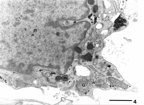 Fig. 4 : A high magnification micrograph showing the cytoplasmic details of the type I agranular interstitial trophoblast cell. A cluster of glycogen (g), mitochondria (m), Golgi apparatus (G) and secretory granule (s). Nucleus (n).(Mag: x27791, Bar: 1 um).
Immunogold Labelling
Sections labelled for cytokeratin showed a significant staining of the type I [Figure 5] and the type II interstitial trophoblast cells [Figure 6a and 6b]. However, the intensity and distribution of gold labelling varied between the cell profiles. The gold particles on the type I interstitial trophoblast were localised specifically on bundles of filaments, whereas the gold particles on the type II cells appeared to be distributed more generally throughout the cell cytoplasm. Sections immunolabelled for hPL demonstrated a significant amount of labelling on the cytoplasmic granules of both the type I and II cells [Figure 6c].
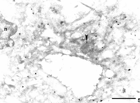 Fig. 5. A type I agranular interstitial trophoblast cell from tissue embedded in Lowicryl HM 20 and immunolabelled with mouse anti-cytokeratin followed by streptavidin-1 nm gold particles. A significant number of gold particles are present on the bundles of filaments in the cytoplasm (arrowhead). Intracellular space (i); Nucleus (n).
(Mag: x27791, Bar: 1 um).
Fig. 5. A type I agranular interstitial trophoblast cell from tissue embedded in Lowicryl HM 20 and immunolabelled with mouse anti-cytokeratin followed by streptavidin-1 nm gold particles. A significant number of gold particles are present on the bundles of filaments in the cytoplasm (arrowhead). Intracellular space (i); Nucleus (n).
(Mag: x27791, Bar: 1 um).
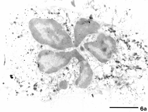 Fig.6a. Electron micrograph of a type II granular interstitial trophoblast cell immunolabelled for cytokeratin. Silver enhanced gold particles are seen over the cytoplasm over individual filaments.
(Mag: x17500, Bar: 2 um).
Fig.6a. Electron micrograph of a type II granular interstitial trophoblast cell immunolabelled for cytokeratin. Silver enhanced gold particles are seen over the cytoplasm over individual filaments.
(Mag: x17500, Bar: 2 um).
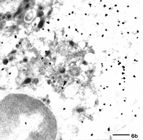 Fig.6b: A higher magnification of another type II cell. Numerous gold particles (arrowhead) are seen over the cytoplasm, bundles of filaments are not seen.(Mag: x21312,Bar: 1 um).
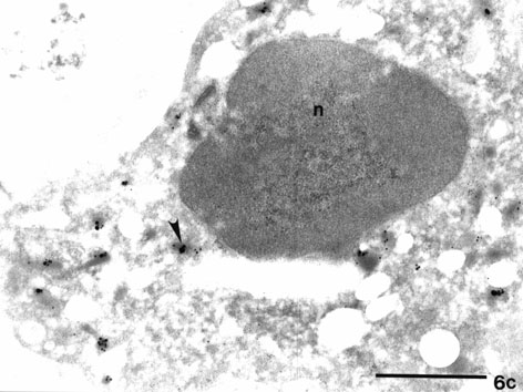 Fig.6c. Electron micrograph from a section immunolabelled for anti-human placental lactogen followed by streptavidin/1 nm gold particles and silver enhancement. Numerous gold particles (arrowhead) are present on the cytoplasmic granules of this type II granular interstitial trophoblast cell.(Mag: x40,300; Bar: 1 um).
.
| <= Materials & Methods | RESULTS | Discussion & Conclussions => |
| Discussion Board | Next Page | Your Poster Session |