 At
these sites, muscle fibers were attached to the bone via the
periosteum. Sharpey's fibers had infiltrated between the periosteum
and the bone, and a layer of membranous ossification was evident at
the site of infiltration.
At
these sites, muscle fibers were attached to the bone via the
periosteum. Sharpey's fibers had infiltrated between the periosteum
and the bone, and a layer of membranous ossification was evident at
the site of infiltration.| INABIS '98 Home Page | Your Poster Session | Related Symposia & Posters | Plenary Sessions | Exhibitors' Foyer | Personal Itinerary | New Search |
1.Histological examination of the control group
The attachment site of the deep layer of the masseter muscle to the mandible was classified into three sites based on the morphology of the attachment to the bone5,6): the periosteal attachment site, transition site, and tendinous attachment site.
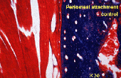
1)Periosteal attachment site (Fig.2)
At
these sites, muscle fibers were attached to the bone via the
periosteum. Sharpey's fibers had infiltrated between the periosteum
and the bone, and a layer of membranous ossification was evident at
the site of infiltration.
2)Transition site
t these sites, the morphology of attachment suggested a transition from periosteal to tendinous attachment sites.
3)Tendinous attachment site
At these sites, muscle fibers were attached to the tendon. The tendon was connected to the bone tissue.
2.Histological examination of the experimental groups
1)Three days after surgery (Fig.3)
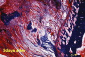 The
tendinous attachment site and the transition site were damaged and
had lost its alignment. The muscle fibers of the periosteal
attachment site were severed and had lost continuity. Similar
findings were observed in the other specimens of this group. These
findings made little difference compared to the ones observed in
seven days after surgery.
The
tendinous attachment site and the transition site were damaged and
had lost its alignment. The muscle fibers of the periosteal
attachment site were severed and had lost continuity. Similar
findings were observed in the other specimens of this group. These
findings made little difference compared to the ones observed in
seven days after surgery.
2) Fourteen days after surgery (Fig.4)
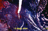 The
tendinous tissue ( the outer most layer of the masseter musle ) of
the tendinous attachment site had been repaired and had regained its
alignment. However, the periosteal attachment site and the transition
site had not yet recovered. By this time, the rats were not only soft
but also regular feed, suggesting that their masticating function had
recovered to some degree. Similar findings were observed in the other
specimens of this group.
The
tendinous tissue ( the outer most layer of the masseter musle ) of
the tendinous attachment site had been repaired and had regained its
alignment. However, the periosteal attachment site and the transition
site had not yet recovered. By this time, the rats were not only soft
but also regular feed, suggesting that their masticating function had
recovered to some degree. Similar findings were observed in the other
specimens of this group.