Pharmacology & Toxicology Poster Session
| INABIS '98 Home Page | Your Session | Symposia & Poster Sessions | Plenary Sessions | Exhibitors' Foyer | Personal Itinerary | New Search |
Introduction
Beneficial effects of marine oil diets containing high level of n-3 PUFAs on cardiovascular and inflammatory diseases are well documented (Schmidt, 1997). In contrast increasing number of papers appear demonstrating undesired effects of n-PUFAs consumption. In our previous work suppressive effect of n-3 PUFA rich diet on the reparative regeneration of the connective tissue was shown (Arend et al., 1996). Feeding rats for three weeks with diet enriched with n-3 fatty acids decreased content of the 2-series PGs, increased the basal level of lipid peroxidation and their peroxidizability in the proliferating connective tissue of the liver wound. Elevated levels of lipid peroxidation products may be one of the reasons for shown suppression of wound healing. In the present study antioxidative treatment with alpha-lipoic acid was performed. alpha-Lipoic acid both as an metabolic antioxidant and a drug has in recent years gained considerable attention (Packer et al., 1998). Its therapeutic effect has been related to the antioxidant activity connected with the ability to scavenge reactive oxygen. It also protects membranes by interacting with vitamin C and glutathione, which may in turn recycle vitamin E and has ability to repair oxidative damage.
Materials and Methods
Experiments were performed on male albino Wistar rats (weight 220-250 g). Animals were given commercial rat pellet diet (control group) or diets enriched with 10% of sunflower oil (n-6 group) or 10% of fish oil (n-3 group) for 8 weeks. After that alpha-lipoic acid (DL-6,8-thioctic acid [Sigma]) was added to the same diets at the calculation of 30 mg per rat for 10 days before the liver thermic wound was induced. The wounds were made after laparotomy by the application of the galvanic cauter's needle to the surface of the liver. Starting from Day 3 and especially on Day 6 the proliferating connective tissue forms a zone distinctly delimited from the central necrotic focus from one side and the hepatic parenchyma from the other side, which allows easy and distinct histomorphological measurements of the width of the connective tissue zone as can be seen in Figure 1.
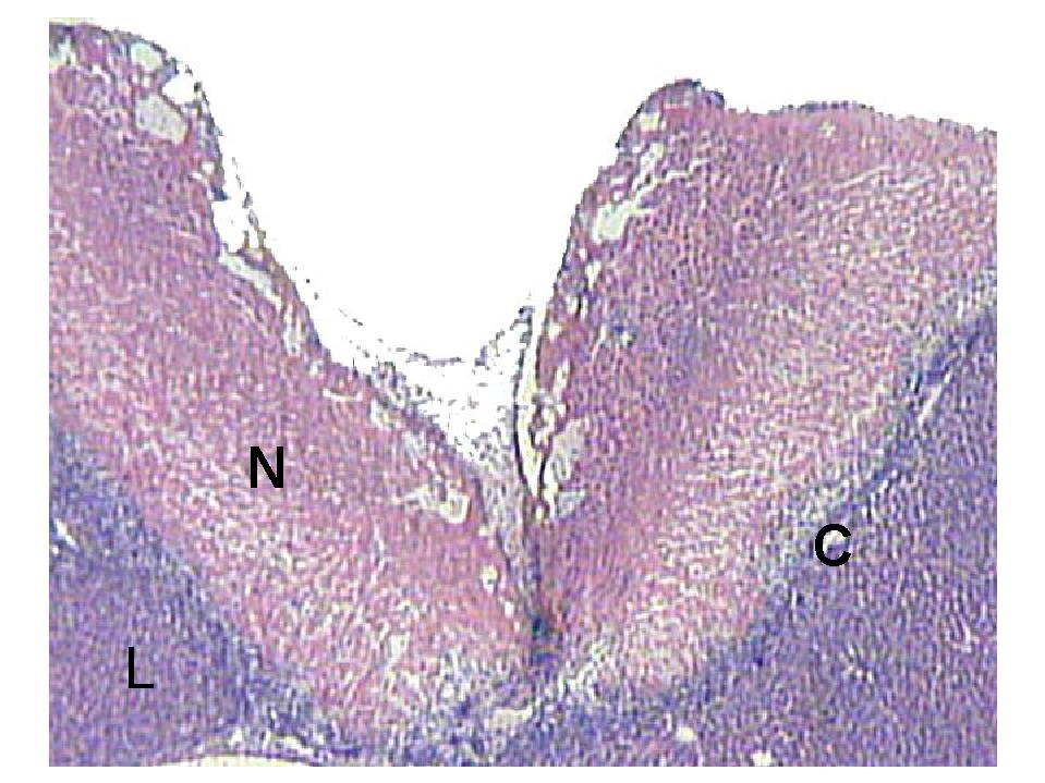 Fig. 1: Panoramic view of the liver wound. L = liver ; C = connective tissue; N = necrosis.
Lipoic acid treatment was continued after wound infliction for 6 days, then in the liver wound the proliferation of the connective tissue (width of the connective tissue zone), the level of lipid peroxidation (TBARS) and peroxidizability (Fe-TBARS), and the content of prostaglandin E2 and F2alpha (radioimmunoassay) was estimated.
In a separate experiment lipoic acid (30 mg/rat) was added to the standard diet for 10 days, then in the liver tissue the level of oxidized and reduced glutathione (GSSG and GSH, respectively) was measured.
Results
Both diets (either rich in n-6 or in n-3 polyunsaturated fatty acids) in case of lipoic acid administration did not affect the wound healing as no changes were seen in the width of the connective tissue zone in the liver wound. Similarly the basal level of lipid peroxidation (TBARS) remained unchanged. Still the peroxidizability of lipids (Fe-TBARS) was slightly elevated in n-3 group as compared to control or n-6 group as seen in Figure 2.
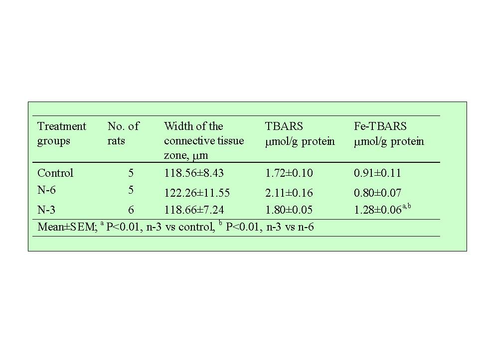 Fig. 2: Effects of diets on the connective tissue proliferation (width of the connective tissue zone) and on the level of lipid peroxidation (TBARS) and on the peroxidizability of lipids (Fe-TBARS) in the liver wounds on Day 6 in the presence of lipoic.
No differences were seen in content of prostaglandins between n-6 and control groups. At the same time the diet rich in n-3 PUFAs significantly decreased the level of prostaglandin E2 and F2alpha in the liver wound and in the liver tissue as shown in Figure 3.
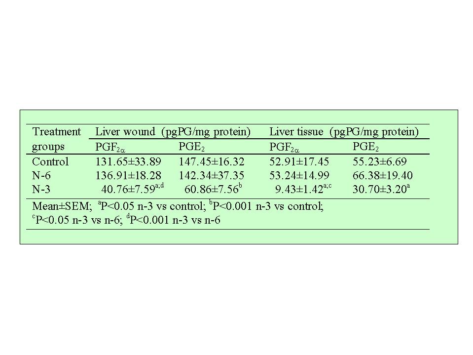 Fig. 3. Effects of diets on the content of prostaglandins (PG) in the liver wound and in the liver tissue on Day 6 in the presence of lipoic acid.
Measurement of gluthatione in the liver tissue demonstrated the effect of lipoic acid to decrease the level of GSSH and to increase the level of GSH thus significantly improving the GSSG/GSH ratio (0,53�0,01 in control vs 0,28�0,04 after lipoic acid administration).
Discussion and Conclusion
As shown in our previous study (Arend et al., 1996) the n-3 PUFAs rich diet enhance the lipid peroxidation in rat liver and suppress the liver wound healing as seen in Figure 4.
 Fig. 4: Effect of lipoic acid on the connective tissue proliferation (wound healing), on the lipid peroxidation (TBARS), on the peroxidizability of lipids (Fe-TBARS), and on the level of prostaglandins (PGF2alpha and PGE2) in the 6-day liver wound of rats (6 per group).
Increased lipid peroxidation and rapidly altered spectrum and/or decreased level of 2-series PGs are evidently the reasons for suppression of the connective tissue proliferation by fish oil. Due to the facts that several products of lipid peroxidation are potent chemoatractants the normal migration of monocytes and proliferation of fibroblasts is disturbed.
Considering several well-documented beneficial effects of lipoic acid, a metabolic antioxidant, we examined the effect of administration of lipoic acid on wound healing in case of n-3 and n-6 rich diet. Our experiments demonstrate the ability of lipoic acid to avoid the adverse effect (suppression of wound healing) caused by consumption of n-3 PUFAs. The positive potency of lipoic acid becomes especially clear if to compare the results of this and previous study (Fig 4). One of the obvious effects of alpha-lipoic acid administration is related to its ability to prevent the lipid peroxidation enhanced by n-3 fatty acids diet. Although the peroxidizability of lipids, caused by n-3 fatty acids diet, remains slightly elevated even after the lipoic acid administration, the significantly lower range of peroxidizability of lipids is quite obvious if to compare the experiments with and without lipoic acid (Fig. 4).
As previously shown n-3 PUFAs administration suppress the formation of 2-series PGs. Evidently it is caused by inhibiting action of n-3 PUFAs on arachidonic acid metabolism yielding in rise of the 3-series PGs and 5-series leukotrienes. As compared to arachidonic acid derivatives these eicosanoid have in general lower activity (Galli et al., 1993). In the present study we established that the administration of alpha-lipoic acid abolished to a certain extent strong decreasing effect of n-3 PUFAs diet on content of 2-series PGs. Although the administration of lipoic acid was unable to fully recover the n-3 fatty acids caused suppression of the 2-series PGs, the normalizing influence is obvious. The effect of lipoic acid to minimize the decrease in the 2-series PGs becomes especially clear if to compare our two studies (Fig 4). Despite the ineffectiveness of lipoic acid administration to fully maintain PGs at the usual level, the induced changes in the prostaglandin spectrum are sufficient for normal inflammatory response and normal wound healing.
In conclusion, feeding rats with diet significantly enriched with n-3 PUFAs suppress reparative regeneration of the connective tissue in rat liver wound. The preventive influence of lipoic acid on n-3 PUFAs induced suppression of wound healing is caused by several actions like prevention of increased (abnormal) lipid peroxidation, and maintaining of sufficient spectrum and/or level of PGs. The crucial protective effect of lipoic acid is obviously achieved by elevation of the content of glutathione, known as important cellular antioxidant, in liver by elevating GSH and decreasing GSSH.
References
- Arend, A; Zilmer, M; Zilmer, K (1996) Dietary n-3 polyunsaturated fatty acids suppress reparative regeneration of the rat liver connective tissue. In: Kumpulainen, J.T.; Salonen, J.T., eds. Natural Antioxidants and Food Quality in Atherosclerosis and Cancer Prevention. Oxford, Thomas Graham House, 60-66.
- Schmidt, E.B (1997) n-3 fatty acids and the risk of coronary heart disease. Dan Med Bull, 44:1-22.
- Packer, L (1998) alpha-Lipoic acid: a meatbolic antioxidant which regulates NF-kB signal transduction and protects against oxidative injury. Drug Metabolism Reviews, 30:245-275.
- Galli, C; Marangoni, F; Galella, G (1993) Modulation of lipid derived mediators by polyunsaturated fatty acids. Prostaglandins Leukotrienes and Essential Fatty Acids, 48:51-55.
| Discussion Board | Previous Page | Your Poster Session |