Medicine Poster Session
| INABIS '98 Home Page | Your Session | Symposia & Poster Sessions | Plenary Sessions | Exhibitors' Foyer | Personal Itinerary | New Search |
Introduction
In a normal liver, populations of parenchymal hepatocytes and nonparenchymal cells are maintained in a constant ratio, and the synthesis and degradation of extracellular matrix components are well balanced. However, homeostasis of the interaction between hepatic cell populations and the matrix metabolism is disturbed by viral infections or by chronic hepatic injury resulting from continuous exposure to toxic chemicals and excessive alcohol consumption. Consequently, extracellular matrix components accumulate in the liver, and nonparenchymal septal cells, including transformed stellate cells (myofibroblasts), increase in fibrous regions (1-3). This chronic liver disease is known as liver cirrhosis, and is accompanied by hypo-albuminemia, ascites, and esophageal varices. Severe hepatic failure, hepatocellular carcinoma (HCC), and/or encephalopathy are induced in the later stage of the disease.
Treatment for liver cirrhosis is not yet well established. Hence, research has focused on factors regulating the activation of stellate cells during chronic liver diseases. A number of cytokines, including transforming growth factor beta 1 (TGFbeta1) (2,4,5) and platelet-derived growth factor (PDGF) (2,6,7), have been suggested to be involved in the up-regulation of the fibroblast-like cells. These findings prompted us to establish gene therapy for the cirrhotic liver by delivery of a gene that would block or counteract the over-production of extracellular fibrous components. One of the greatest hurdles is to develop an appropriate strategy to target exogenous gene delivery to the septal cells in the cirrhotic liver.
It is well known that adenovirus infects almost all hepatocytes through the blood circulation from the portal vein, peripheral vein or abdominal cavity of normal rats (10,11). Therefore, in this study, we examined whether recombinant adenovirus vectors could transfer a foreign gene to the septal cells of rat livers with cirrhosis induced by carbon tetrachloride (CCl4).
Materials and Methods
Animals
Thirty male Wistar rats weighing 150 g were purchased from Shizuoka Laboratory Animal Center (Japan). Liver cirrhosis was induced by intraperitoneal injection with 1 ml of CCl4: olive oil (1:1 mixture) per kg of body weight, twice a week for 4-8 weeks, as described previously (12,13). All rats were fed ad libitum, and received humane care in compliance with the institution's guidelines for the care and use of laboratory animals in research.
Culture of liver slice
Liver slices were prepared and cultured basically as described previously (14,15). Briefly, the liver lobes of the cirrhotic rats were quickly removed and placed in Dulbecco-modified MEM supplemented with 5.6 mM D-glucose, 26 mM NaHCO3, 2 mM glutamine, 10% fetal calf serum, 50 mg/l streptomycin and 5X10⁴ units/l penicillin. Small pieces of the liver were sliced into 100-um sections with a microslicer (Dosaka EM Co., Ltd., Tokyo, Japan). The sections were cultured in 5% CO2 at 37Celcius in the medium mentioned above.
Infection of the adenovirus in vivo and in vitro
Normal, fibrotic and cirrhotic rats were infected with 100 ul (1.5X10⁹ pfu) of the purified recombinant adenovirus Adex1CALacZ via the tail vein. The infected rats were sacrificed at 4 days post-infection, and the livers or kidneys were fixed by perfusion from the heart with chilled phosphate-buffered saline (PBS) followed by 2% formaldehyde, 0.2% glutaldehyde and 0.1% NP40 in PBS. These tissues were sliced into 10-um sections with a cryostat. The expression of beta-galactosidase was determined in these sections on the glass slides.
Slice cultures of the cirrhotic livers were infected with the vectors (1X10⁶ pfu), and incubated in 5% CO2 at 37Celcius for 24 h. The slices were fixed with formaldehyde solution as described above, and stained with X-gal.
Beta-galactosidase histochemical assay
Determination of the expression of the lacZ gene was carried out according to Jaffe et al. and Bao et al. (10,16). Briefly, the fixed specimens were rinsed three times with PBS and incubated in a reaction mixture containing 5 mM K3Fe(CN)6, 5 mM K4Fe(CN)6, 2 mM MgCl2 and 1 mg/ml X-gal (5-bromo-4-chloro-3-indolyl-beta-D-galactopyranoside) in PBS for 2 h at 37Celcius. Subsequently, these specimens were counterstained with eosin, or with anti-vimentin antibody.
Results
In normal rat livers, almost all hepatocytes expressed beta-galactosidase from the recombinant adenovirus vectors (Fig. 1a). In cirrhosis, there is an even stronger reduction in the number of transduced hepatocytes (Fig. 1b). On the other hand, in vitro infection to the slice culture demonstrated that the cirrhotic hepatocytes still maintained adenovirus infectivity (Fig. 2a). These results showed that the hepatocytes of the cirrhotic liver were susceptible to adenovirus infection, but that the septal cells are likely to create a barrier to in vivo infection of hepatocytes in cirrhotic livers. Moreover, in the kidney, which is the second target organ of adenovirus, there was no difference in infectivity between normal and cirrhotic rats (Fig. 2b).
The recombinant adenovirus intravenously transmitted the foreign gene to septal cells in extranodular fibrous septa, rather than to hepatocytes within small nodules. Therefore, septal cells in the cirrhotic liver should be targeted with the adenovirus vector for a successful in vivo therapeutic strategy.
Fig.1a
|
Fig.1b
|
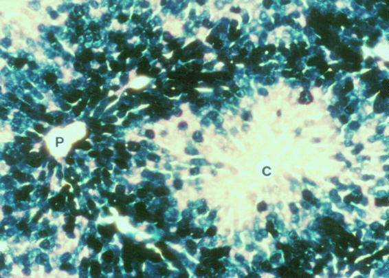
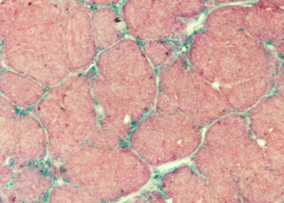
- Fig. 1. Expression of the lacZ gene in normal, fibrotic and cirrhotic rat livers. In the normal liver (a), numerous hepatocytes show positive reaction with X-gal staining. P: portal vein, C: central vein. In severe cirrhotic liver (b), X-gal positive cells are observed in septal cells in the connective tissues of fibrous septa.. Magnification is X80 (a); X33 (b).
Fig.2a
|
Fig.2b
|

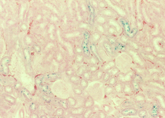
- Fig. 2. Infectivity in liver and kidney cells of the cirrhotic rat. In the cirrhotic liver slice (a) infected with the vector in vitro, X-gal positive cells are observed in both hepatocytes (arrowheads) and septal cells (arrows) in fibrous septa in cirrhosis. Kidneys in the cirrhotic rat (b) were examined for virus infectivity in vivo. Blue stained cells by X-gal show infected cells. Magnification is X90 (a); X70 (b).
Discussion and Conclusion
In the present study, we attempted to target the septal cells in the cirrhotic liver with the adenoviral gene delivery system. Our results demonstrated that hematogenous infection of the recombinant adenovirus selectively transmitted the foreign gene to the septal cells in the extranodular fibrous septa, rather than to hepatocytes within small nodules, with no increase of infection into the kidney as the second target organ of adenovirus (11).
Although parenchymal cells in cirrhosis still upheld susceptibility to the adenovirus vector, as shown by the slice culture experiment, the hepatocytes within small nodules in cirrhotic liver scarcely exhibit adenovirus-mediated in vivo gene expression. Taken together with the observation that intralobular hemodynamics is reduced by the formation of shunts between portal and central veins, the shunting of blood away from the hepatocytes may be an important factor which could lead to preferential transfection of septal cells. On the other hand, adenovirus infection depends on attachment of a viral fiber to coxsackie/adenovirus receptor (CAR) in target cells (17, 18), and following internalization by endocytosis-mediated integrins (19). Both CAR and ALPHAv integrins have been reported to be highly expressed in the normal murine liver (18, 20); however, little is known about the expression of CAR and integrins in cirrhotic liver. The inefficient gene transfer to hepatocytes in cirrhosis may be related to decreasing levels of CAR and/or integrins, as well as to shunting of the blood flow.
References
- Milani S, Herbst H, Schuppan D, Hahn EG, Stein H. Hepatology 1989; 10: 84-92.
- Gressner AM. Z Gastroenterol 1992; (Suppl.1): 5-16.
- Seifert WF, Roholl PJ, Blauw B, van-der-Ham F, van Thiel De Ruiter CF, et al. Int J Exp Pathol 1994; 75:131-46.
- Czaja MJ, Weiner FR, Flanders KC, Giambrone MA, Wind R, Biempica L, et al. J Cell Biol 1989; 108: 2477-82.
- Sun MA, Wang BE, Annoni G, Degli Esposti S, Biempica L, Zern MA. A morphologic and molecular comparison. Lab Invest 1990; 63: 467-75.
- Friedman SL, Arthur MJ. J Clin Invest 1989; 84: 1780-5.
- Pinzani M, Gesualdo L, Sabbah GM, Abboud HE. J Clin Invest 1989; 84: 1786-93.
- Matsuda Y, Matsumoto K, Yamada A, Ichida T, Asakura H, Komoriya Y, et al. Hepatology 1997; 26: 81-89.
- Yasuda H, Imai E, Shiota A, Fujise N, Morinaga T, Higashio K. Hepatology 1996; 24: 636-42.
- Jaffe HA, Danel C, Longenecker G, Metzger M, Setoguchi Y, Rosenfeld MA, et al. Nat Genet 1992;1: 372-8.
- Huard J, Lochmuller H, Acsadi G, Jani A, Massie B, Karpati G. Gene Ther 1995; 2: 107-15.
- Hirayama C, Morotomi I, Hiroshige K. Biochem J 1970; 118: 229-32
- Ala Kokko L, Stenback F, Ryhanen L. Biochem J 1987; 246: 503-9.
- Hart A, Mattheyse FJ. In Vitro 1983; 19: 841-52.
- Balinsky JB. Bhattacharya R, Rao PV, Bhaskar AS, Pant SC, Dube SN. Hum Exp Toxicol 1996; 15: 105-10.
- Bao JJ, Zhang WW, Kuo MT. Hum Gene Ther 1996; 7: 355-65.
- Bergelson JM, Cunningham JA, Droguett G, Kurt Jones EA, Krithivas A, Hong JS, et al. Science 1997; 275: 1320-3.
- Bergelson JM, Krithivas A, Celi L, Droguett G, Horwitz MS, Wickham T, et al. J Virol 1998; 72: 415-9.
- Wickham TJ, Mathias P, Cheresh DA, Nemerow GR. Cell 1993; 73: 309-19.
- Huard J, Lochmuller H, Acsadi G, Jani A, Holland P, Guerin C, et al. Exp Mol Pathol 1995; 62: 131-43.
| Discussion Board | Previous Page | Your Poster Session |