Cardiovascular Diseases Poster Session
| INABIS '98 Home Page | Your Session | Symposia & Poster Sessions | Plenary Sessions | Exhibitors' Foyer | Personal Itinerary | New Search |
Materials and Methods
The period of this study was from May 1990 to December 1996 on a total of thirty-six patients. [28 males and 8 females]. Angiography showed five aorotoiliac and twelve mid-femoral blocks. Thirteen had blocks in the anterior/posterior tibial or peroneal arteries. Raynaud’s cases had no block in major vessels but small artery blocks with gangrenous finger tips. Twenty-two cases presumably had thromboangitis obliterance [young chronic smokers with an extensive block], five had atherosclerosis [older patients with patchy blocks], and in three cases vascular disease was accompanied by diabetes mellitus, and six had Raynaud’s in upper extremities (pulsatile major vessels, blanching, cyanosis. All lower extremity group patients were chronic smokers and their presenting symptoms and signs included severe rest pain. These patients could barely walk because of pain and had cold pale extremities and minor distal gangrenous changes [either toe/s or skin patchRecurrent trauma in barefoot patients, probably, made them more prone to distal gangrenous tips even in patients with higher level arterial blocks. All patients were previously treated with conservative management (pentoxiphyline, analgesics, exercise program ,abstinence from smoking etc). Six patients had failed lumbar sympathectomy and three had failed vascular bypass. Angiography, vascular doppler and oxymetry were routinely carried out in all cases pre and postoperatively.
Operative technique: All operations were done under general anesthesia. Tourniquet was not used for obvious reasons. Through small multiple incisions over the anterior border of the tibia,
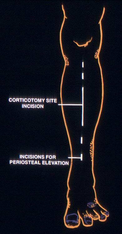 Sites of Incisions
the periosteum was stripped over the medial and lateral surfaces from tibial tubercle to the medial malleolus except at the site of corticotomy.
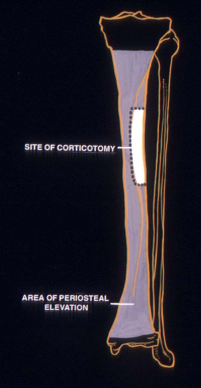 site of corticotomy and area of periosteal elevation.
The corticotomy was performed at a distance of about 10 cms from the knee joint on lateral surface of tibia ,five cms in length and near both anterior and posterior neurovascular bundles. A five cms long incision, parallel and just lateral to the anterior border of tibia was made at the corticotomy site and elevating skin, one cms width of medial surface of tibia is visualized. A guide wire was passed along the antero-lateral surface of the tibia to judge the direction of the drill bit. Five to six drill holes spaced a centimeter apart were made from the antero-medial to the posterior cortex along the endosteal surface.
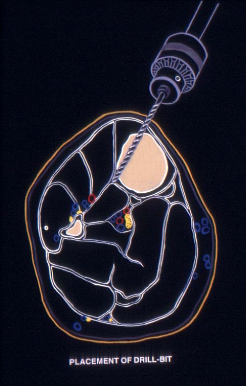 Direction of drillbit at the crossection of leg, 10 cms below the knee joint.
A three-sided corticotomy was then done leaving the posterior side intact. This was manually broken like a hinge using the posterior periosteum as the intact bridge. This corticotomy segment therefore remained adherent to the periosteum and attached to interosseus membrane over which the neurovascular bundles lay. The operation was completed by suturing the wounds, desloughing of distal ulcers and ablating any frankly gangrenous area.
In upper extremity cases, ulna was stripped of the periosteum, and multiple drill holes were placed in ulna and through relevant metacarpals.
Postoperative management: This included mobilization of the knee and the ankle, antibiotics, adequate hydration. Mild analgesics were used but anti-inflammatory drugs were avoided since that conflicts with the principle .No limb elevation or dependency was enforced. Antiplatelet drugs or heparin was not used.
| <= Introduction | MATERIALS & METHODS | Results => |
| Discussion Board | Next Page | Your Poster Session |