
Cancer Poster Session
Abstract
Introduction
Materials & Methods
Results
Discussion & Conclusion
References
Discussion
Board
|
Electrochemotherapy Potentiation of Antitumor Effect of Cyclophosphamide by Local Electric Pulses on the Metastatic Lesion of Hamster Oral Fibrosarcoma
Contact Person: Hatsuhiko Maeda (hatsu@dpc.aichi-gakuin.ac.jp)
Results
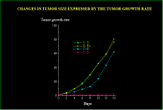
The average size of the hamster oral fibrosarcoma tumor of metastatic lesion was 103 ± 17.80 mm3 at the first day of the treatment. Despite the treatment applied, the tumor of the metastatic lesion grew continuously in 3 groups: D-E- ( non-treatment ), D-E+ ( electric pulses alone ) and D+E- ( drug application alone) groups. However, the D+E+ group which received electric pulses and intraperitoneal injection of CPA, showed a significant reduction in tumor size ( p < 0.001) compared with the other three groups ( Fig. 2). The responses found in D+E+ group were as follows; in the D+E+ group, the average size of the tumor decreased to 41, 30, 19, 31, 64, 133 and 176% of the initial size at 2, 4, 6, 8, 10, 12, and 14th day, respectively, indicating that responses were marked in the early days after the treatment. Furthermore, 1 out of 10 (10%) hamsters showed a complete regression. In contrast, no response ( decrease in tumor size ) was seen in any hamsters in the D-E- , D-E+ and D+E- groups(Fig. 3a, b).
 Click to enlarge
 Fig.2: Changes in tumor size expressed by the tumor growth rate. This growth rate is defined as Vn/V0, where V0 is an initial tumor volume and Vn is the volume at the nth day after treatment. In the D+E+ group, a significant regression of the tumor size is observed(p<0.001).
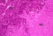
 Click to enlarge
 Fig. 3a: D-E- group(control group), at 14th day. The tumor increased to approximately 7600% of the initial size. D-E+ group( electric pulses alone ) also showed the same figure. NL: normal lymph node, ML: metastatic lesion.
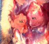
 Click to enlarge
 Fig. 3b: Metastatic lesion of the lymph node. . Tumor cells invaded in the lymph node. NL: normal lymph node, ML: metastatic lesion.
HE x40
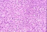
During the experiment, biopsies were taken from the animals of 4 groups on days 0, 2, 4, 8 , 10, 12 and 14, and they were examined histologically. The histological examination revealed that the tumors of the D+E+ groups were highly necrotic ( Fig. 4 ). In contrast, the hamsters of both D-E- and D-E+ groups had well-defined active tumors which consisted of spindle shaped cells with round nuclei, showing sporadic mitotic figures(Fig. 5, 6). In the D+E- group, the mixture of apoptotic or necrotic tumor cells and viable tumor cells were observed (Fig. 7 ). No burns were observed on the oral mucosa of the animals exposed to electric fields. However, these animals showed signs of edema for 1-3 days after the treatment.
 Click to enlarge
 Fig.4: Histological examination of biopsies. Hematoxylin-eosin stains from samples of biopsies taken at day 4: D+E+ group, all tumor cells show necrotic figures. HE x100.
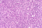 Click to enlarge
 Fig.5: Histological examination of biopsies. Hematoxylin-eosin stains from samples of biopsies taken at day 4: D-E- group(control group), viable tumor cells which are spindle shaped and with round nuclear, show several mitotic figures. HE x100.
 Click to enlarge
 Fig.6: Histological examination of biopsies. Hematoxylin-eosin stains from samples of biopsies taken at day 4: D-E+ group, tumor cells show the feature similar to those of D-E- group(control group) HE x100.
 Click to enlarge
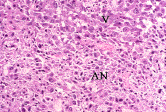
 Fig.7: Histological examination of biopsies. Hematoxylin-eosin stains from samples of biopsies taken at day 4: D+E- group, mixture of apoptotic, necrotic tumor cells (AN) and viable tumor cells (V) are seen HE x100.

AUTHOR'S BRIEF EXPLANATION
(This means EXPLANATION BY AUTHORS)
This study has clearly demonstrated that electrochemotherapy with the injection of cyclophoshamide is an effective treatment for decreasing tumor volume of the metastatic tumor site of the regional lymph node..
Back to the top.
| Discussion Board | Next Page | Your Poster Session |
|
|
Maeda, H;
Kojima, A;
Sugita, Y;
Tanaka, S;
Kubo, K;
Sato, E;
Konishi, S;
Tanaka, H;
Kameyama, Y;
(1998). Electrochemotherapy Potentiation of Antitumor Effect of Cyclophosphamide by Local Electric Pulses on the Metastatic Lesion of Hamster Oral Fibrosarcoma. Presented at INABIS '98 - 5th Internet World Congress on Biomedical Sciences at McMaster University, Canada, Dec 7-16th. Available at URL http://www.mcmaster.ca/inabis98/cancer/maeda0671/index.html
|
 Click to enlarge
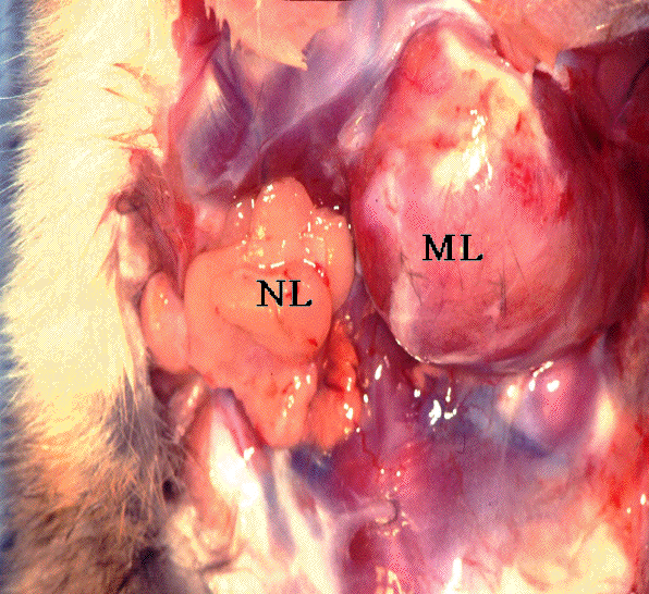 Click to enlarge
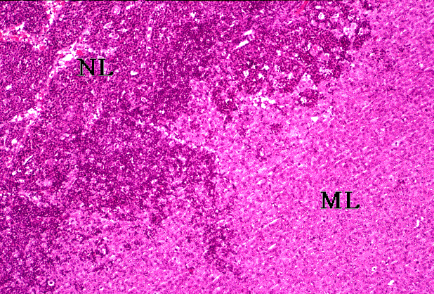 Click to enlarge
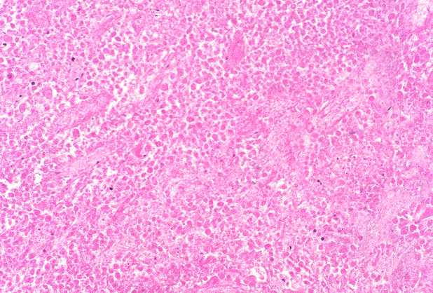 Click to enlarge
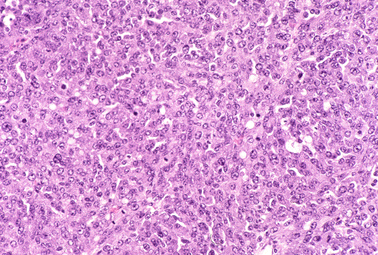 Click to enlarge
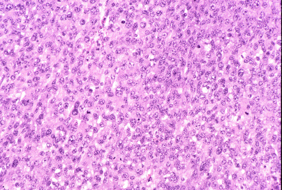 Click to enlarge
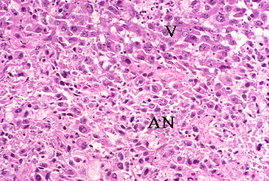 Click to enlarge