Poster Contents
| INABIS '98 Home Page | Your Session | Symposia & Poster Sessions | Plenary Sessions | Exhibitors' Foyer | Personal Itinerary | New Search |
Results
Renal carcinoma cells In the present study, we could obtain limited numbers of the renal carcinoma cells and could observe growth of corresponding autologous lymphocytes as shown in Table I. The carcinoma cell lines from patient series Hpt. and Pt. were established in the developing stage of the primary culture technique in that 7 tumor cell lines grew up out of 38 patients. After the protocol for the routine primary culture (see Materials and Methods) was set during 1995 - 1996, 10 cases were submitted for the primary renal carcinoma cell culture. However, only 3 cell lines were established (patient TUHR series in Table I). The expression of MHC-class I was confirmed in all cases shown in Tabl I except TUHR4TKB by conventional flow cytometric analysis. The positively stained carcinoma cells showed an average of 10-100 fold higher fluorescent intensity than those stained with FITC-labelled control antibodies (data not shown).
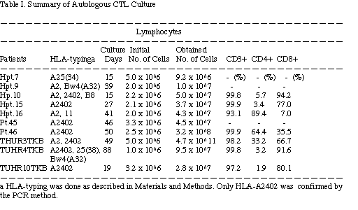
We could also obtain the counterpart normal kidney cells from 5 cases in the former series of patients and only one case, TUHR10TKB, in the latter series. Therefore, to our regret, the autologous full-set (i.e., sufficient number of renal carcinoma cells, normal kidney cells, and PBMC from the same patient for induction of CTL and followed killing assays) was rare. We could examine only 10 cases as shown in Table I including 4 imcomplete sets which lack normal kidney cells because of their short life span (i.e., Hpt.10, Hpt.16, TUHR3TKB, and TUHR4TKB as shown in Table II).
Induction of CTL In a preliminary experiment for induction of allogeneic CTL from PBMC of a healthy volunteer against a gastric carcinoma cell line GT3TKB, effect of different combination of the interleukins was tested. We observed that CTL induced with the four interleukins (IL-1, -2, -4, and -6) showed the stronger killing activity of the target carcinoma cells than those induced with IL-2 only that is widely used in the traditional method for CTL generation (Fig. 1). At an E/T ratio of 1 in the assay course of 24 h, the more interleukins (IL-1, -4 and -6) added in the induction culture produced the more active CTL. Therefore, the IL cocktail of IL-1, -2, -4 and -6 was used in the following experiments of autologous CTL induction against tumor cells.
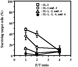
Fig. 1. Effect of the combination of interleukins on the cytotoxci activities of induced CTL. PBMC from a healthy volunteer (2 x 10⁶ cells) were co-cultured with the allogeneic gastric carcinoma cells, GT3TKB, in the medium containing different interleukin combination. The CTL were restimulated with the target cells up to 4 times. After 44 days, the CTL were submitted for the cytotoxicity assay. The killing assay was performed for 24 h at E/T ratios indicated. After the coculture of the effector cells with target carcinoma cells, the lymphocytes were gently washed out, then the adhering (therefore conceivable to be surviving) target carcinoma cells were fixed and stained with 0.4% crystal violet and quantified. CTL were induced with the medium containing various combination of interleukins. Each bar shown is the mean of triplicate observations & plusminus SD.
In the present protocol for induction of autologous CTL with the IL cocktail, lymphocytes in the PBMC aggregated on the carcinoma cells within 3-5 days and began to proliferate. The target carcinoma cells disappeared completely after 7-10 days. After most of the non-proliferating PBMC initially added had died within 5-10 days, the lymphocytes began to burst (except Hpt.7 in Table I), usually after 10-14 days. This pattern of lymphocyte growth was similar to that observed in the case of induction of allogeneic CTL.21) The lymphocytes were restimulated twice with autologous carcinoma cells within the initial 2 weeks. Two typical cumulative growth curves of CTL were shown in Fig. 2. One with the restimulation by autologous carcinoma cells (TUHR3TKB-CTL) and the other without antigen restimulation (Pt.46-CTL). The restimulation apparently boosted CTL growth from the initial 2x10⁶ cells up to more than 1011 cells in 50 days of culture.
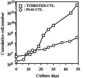
Fig. 2. Typical cumulative grwoth curves of autologous TUHR3TKB-CTL and Pt.46-CTL induced against renal carcinoma cells.
Table I summarizes the autologous CTL induction culture. The phenotypes of the lymphocytes were analysed by flow cytometry except the cases of Hpt.7, Hpt.9, and Pt.45 because of the shortage of yielded lymphocytes. In most cases, the main population of lymphocytes consisted of CD3+CD8+ cells (Table I) except for the cells from patients Hpt.16 and Pt.46. In these cases, CD4+ CTL became dominant after restimulation.
Killing activities of CTL Table II summarizes killing activities and specificity of the autolgous CTL. In 9 out of 10 cases, CTL have been induced. The CTL from patient Hpt.7 lysed the autologous target carcinoma cells at an E/T ratio of 4 in the course of 24 h. Other CTL examined at an E/T ratio of 2 also lysed autologous target cells except in the case of patient TUHR4TKB. The CTL from patients Hpt.15 and Hpt.16 completely lysed autologous target carcinoma cells at this low E/T ratio. However, none of the CTL vigorously killed autologous normal kidney epithelial cells. The CTL from patient TUHR3TKB showed only weak killing activity at this low E/T ratio, and we confirmed that this activity was reproducible. The CTL from patient Pt.46 killed not only autologous target carcinoma cells but also allogeneic Hpt.10 renal carcinoma cells. However, these CTL did not recognize the other renal carcinoma cells tested, OS-RC-2, Hpt.15, TUHR3TKB, and TUHR4TKB.
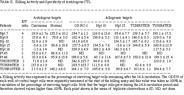
The lymphocytes cultured as CTL from patient TUHR4TKB did not lyse autologous target carcinoma cells, although the major population of cells in this preparation of CTL consisted of CD8+ lymphocytes and these were partially effective in killing the allogeneic TUHR3TKB carcinoma cells. MHC class-I molecules (HLA-A2402) were undetectable on the TUHR4TKB target cell surface (data not shown). In contrast, NK from the same patient (TUHR4TKB) lysed TUHR4TKB carcinoma cells and NK-sensitive K562 cells at an E/T ratio of 2 in the course of 48 h (Fig. 3), but these cells did not lyse other MHC class-I-expressing allogeneic tumor cell lines, TUHR3TKB, Hpt.10, Hpt.15, and OS-RC-2. These data are consistent with the mechanisms of NK recognition and killing.28)
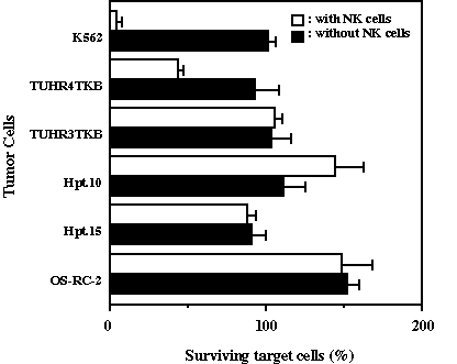
Fig. 3. Cytotoxicity of NK cells towards renal cell carcinomas. Target tumor cells (1x10⁴ cells/well) and NK cells (CD56+ cells were more than 95%) were co-cultured for 48 h at an E/T ratio of 2. The surviving tumor cells were measured as described in Materials and Methods. Note that OD570 of the target carcinoma cells at the start of the coculture was taken as 100%. Also note that the carcinoma cells cultured at the E/T of 0 grew for the 24 h incubation and, therefore, showed more than 100% of surviving. Each bar shown is the mean of triplicate observations & plusminus SD.
The CTL from patient Hpt.9 showed more than 10 fold greater killing activity than autologous LAK and NK (Fig. 4). In other patients, the NK and LAK showed no killing activity, or very limited activity if any, at an E/T ratio of 10 against autologous carcinoma cells (data not shown).

Fig. 4. A comparison of the activity of autologous effector lymphocytes derived from the patient Hpt.9. The target tumor cells and the effector cells were co-cultured for 48 h.
| <= Materials & Methods | RESULTS | Discussion & Conclussions => |
| Discussion Board | Next Page | Your Poster Session |