Biomedical Education Poster Session
| INABIS '98 Home Page | Your Session | Symposia & Poster Sessions | Plenary Sessions | Exhibitors' Foyer | Personal Itinerary | New Search |
Introduction
Attempts have been made to introduce the correlations of general pathology with gross anatomy, as well as clinical relevance, to first year medical students. Historically, such attempts had limitations. They were confined by the available media, and preparation consumed a major amount of time and effort. For example, conventional photographs were taken of the subject matter, often times converted into drawings, and annotated by hand, according to physician’s notes and hospital records. The resulting photographs anddrawings were then assembled into posters, and presented by the students themselves. A significant amount of space was required for the presentation of such posters.
Despite these limitations, we believe that the gross anatomy laboratory provides an excellent opportunity to establish connections between anatomy, histology, and pathology. The intent of this project was to correlate pathology observed during gross anatomy lab, with the relevant anatomy and histopathology, and then present such correlations as self-instructional units to medical students, in order to increase interest and facilitate learning. Current technological advances, specifically in the areas of digital imaging, editing and computerization, have made the aims of this project more readily achievable. The following example is a unit correlating the anatomy, histology, and pathology involved in smoking and lung cancer.
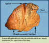
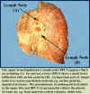
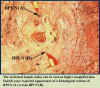
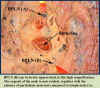
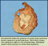
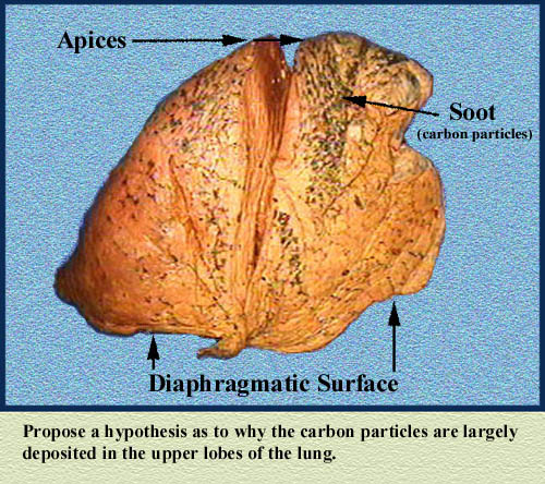
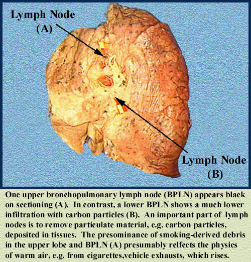
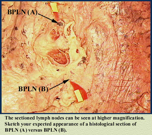
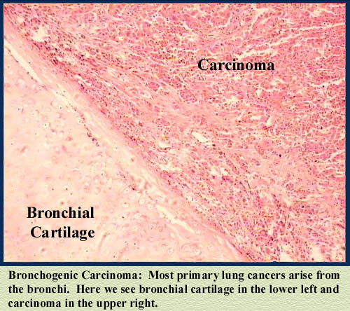
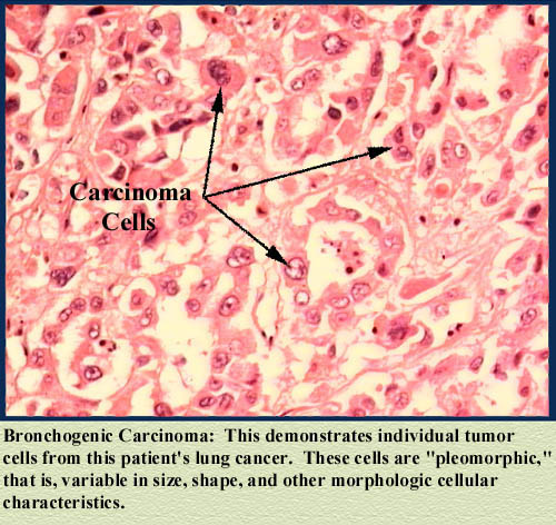
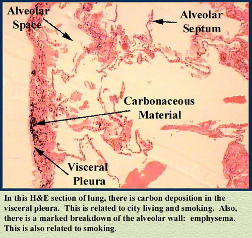
Figure 1 Figure 2 Figure 3 Figure 4 Figure 5 Figure 6
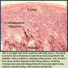
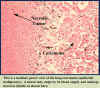
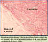
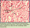
Figure 7 Figure 8 Figure 9 Figure 10 Figure 11

Materials and Methods
The materials and methods evolved during the course of this project. Changes were implemented in order to improve efficiency with respect to time usage and image file size. Initially, a Sony DCR-VX1000 3CCD-System digital video camera was used to record anomalous findings during gross dissections. Still images were then obtained by freezing desired frames, saving them on the computer hard drive, and subsequently onto a 3.5" floppy disk. However, this procedure had limitations. Obtaining adequately lit images throughout the gross anatomy laboratory was cumbersome. Converting the video into still frames via the computer, and then saving to disks became routinely monotonous.
Also, these images could only be saved as large bit-mapped files; therefore, they required an inordinate amount of storage space. As a result of these problems, we decided to record images with the Sony Mavica, MVC-FD7, a digital still camera which uses a 3.5" floppy disk, and is equipped with a 10X zoom lens. This change significantly increased the efficiency of recording images in the gross lab. The advantages of this system were that digital images were recorded directly onto a disk, and because the Mavica saves images as smaller jpeg files, file size was greatly reduced.
A disadvantage was lack of a good three dimensional presentation. Recording of the histopathology images was accomplished with Microimage Video Systems’ AutomatiCam, which was attached to both an inverted Nikon microscope and PC, which also saved the images as jpeg files. Once still images of the gross anatomy and histopathology images were obtained, Adobe Photoshop 5.0 was used to correct for color imbalance, improve clarity and detail, and annotate each image accordingly. These images were imported in a logical sequence into a PowerPoint presentation, and made available for the medical students to review. A survey was used to collect student feedback with regard to interest, understanding, organization, quality, depth, length, and comments.
Results
Results and Discussion
The change in photographic systems made this project much more time efficient and convenient. This was due largely in part on the ability to save images directly on a 3.5" floppy disk as small jpeg files. It should be noted that this system does produce still images of slightly lesser quality; however, unless the image dimensions are expanded beyond reasonable limits, such subtle differences were hard for the human eye to detect.Survey results indicated that 100% of the students surveyed said this unit increased their interest in learning the correlations between gross anatomy, histology, and pathology. Results also indicated that the unit increased student understanding of the material, and they suggested that similar units be made available. Information ascertained regarding the organization, quality, depth, and length of the unit was positive, and on average, rated as "very good." Student comments requested that unit length and depth be increased.
Based on this initial positive reception of the material, the intent is to assemble additional self-instructional "Pathology in the Gross Anatomy Laboratory" units. These units will be accessible to students in a number of ways, including PowerPoint presentations, the intranet, and the internet.

| Discussion Board | Previous Page | Your Poster Session |