He was followed by his general practicioner
He was referred to an orthopaedic Surgeon
Internal Fixation and bone grafting was done on 27th Feb 1991
The bone was sufficiently abnormal for the surgeon to take a specimen for pathological examination.
Case History
The patient is a 57 year old right handed male self employed drywaller. Date of birth 7 May 1941Injury 2 Dec 1990. Age 49 at the time of the index injury.
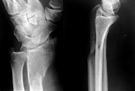
The index injury was an innocuous looking distal radius fracture after fall on the outstretched hand.
|
This was treated by the Emergency Room Physician with a forearm cast. He was followed by his general practicioner |
|
|
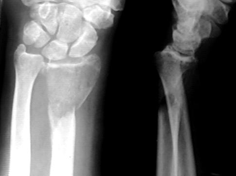
By Feb 25th 1991 the fracture was not healing, had shortened and
angulated. He was referred to an orthopaedic Surgeon Internal Fixation and bone grafting was done on 27th Feb 1991 |
|
|
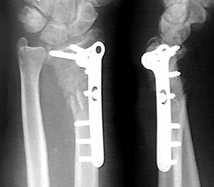
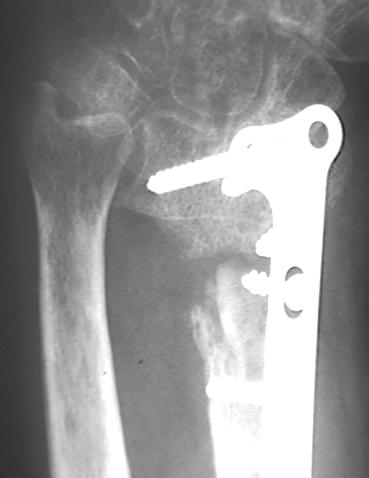
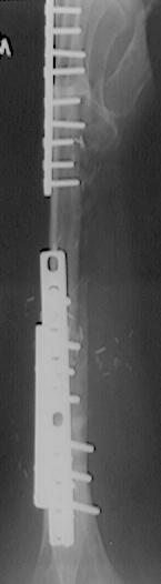
Xray taken 7th March 1991 shows the fracture aligned, bone grafted and
fixed with a T plate The bone was sufficiently abnormal for the surgeon to take a specimen for pathological examination. |
|
|
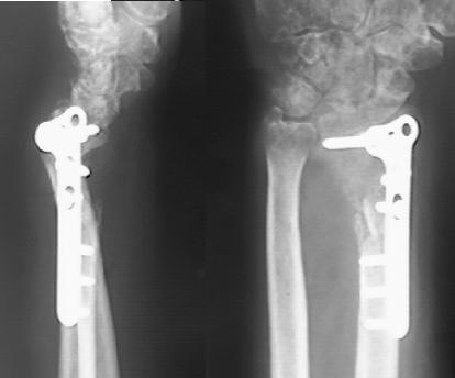
By 13th June 1991 it was noted that the fixation was not holding and the fracture began to displace. | |
 |
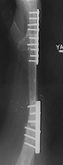
Xray of 28th October 1991 showed significant loss of bone at the non-union site. The bone graft is reabsorbed or fragmenting and there is now significant osteopaenia in the surrounding bone, notably the ulna and the carpal bones. |
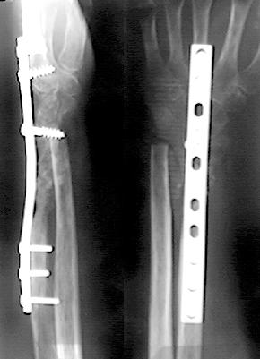 |
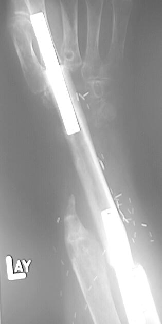
Fusion from the Long finger metacarpal to the radius wilth
resection of ulnar head was undertaken Jan 1992 (L) However, further views taken 10th July 1992 showed reabsorption of bone from the distal ulna and no healing of the fusion. (R) |
 |
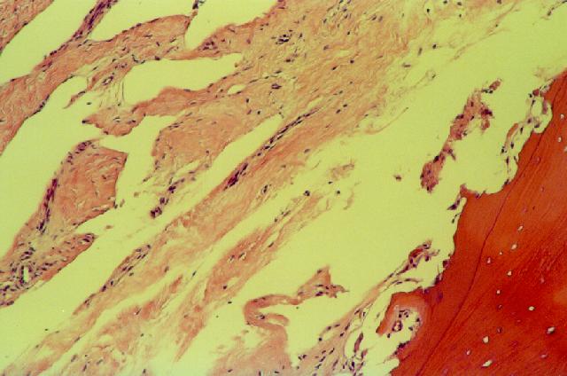 |
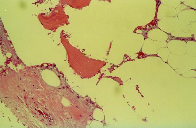
Biopsy of distal L ulna 1993. Ectatic vessels involving the periosteum and adjacent soft tissue. Consistent with but not pathognomonic of the diagnosis of Gorham's Disease |
He was managed non-operatively for 3 years during which time he did return to work as a dry-waller using his right hand only. In 1994 he was referred to a tertiary centre for consideration of vascularized fibular graft. A further biopsy was undertaken and the diagnosis of Gorham/Stout Disease (Disappearing Bone Disease) was made.
 |
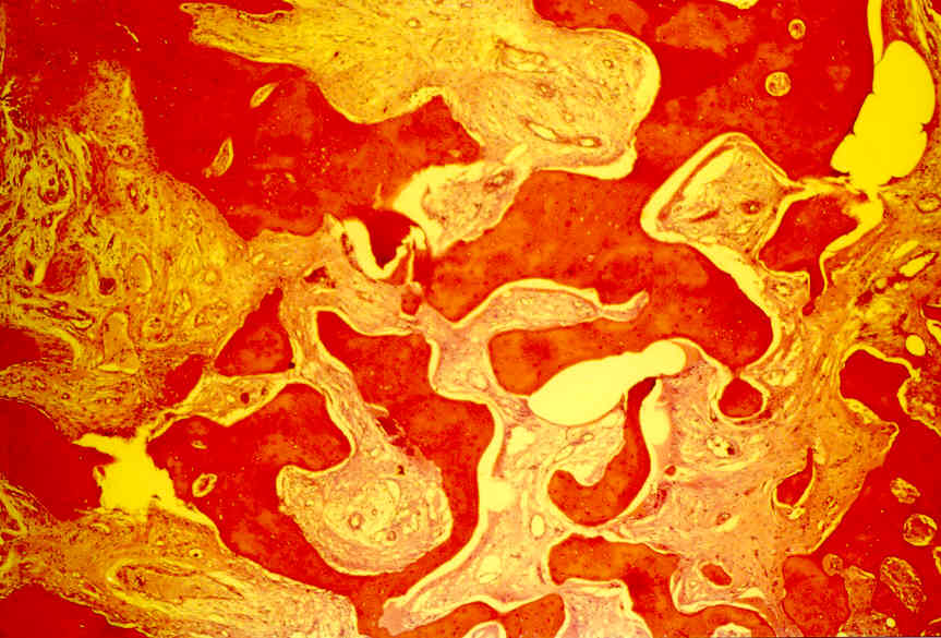
Biopsy L forearm 1994 Cortical and cancellous bone showing osteoporosis with an increased number of capillaries and small arterioles. Consistent with Gorham's Disease |
 |
 |
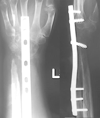
Free vascularized fibular grafting was undertaken bridging from the proximal ulna to the index finger metacarpal. These Xrays from March 27th 1995 show the extent of the bone loss of the radius and carpal bones |
 |
Biopsy L Ulna 1995 Thick and irregularly shaped trabeculae of bone showing focal areas of necrosis and marrow fibrosis |
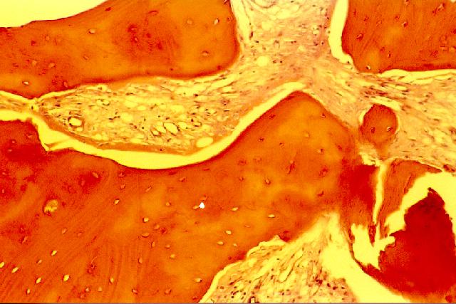 |
Biopsy L Ulna 1995 detail |
 |
Two further procedures were undertaken, application of a
second plate to the proximal site between the ulna and the fibular graft and radiotherapy.
The patient, at least, attributes the eventual healing of the injury to radiotherapy. He
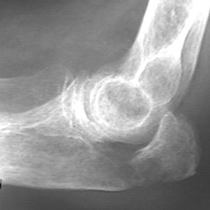
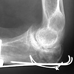
returned to work once again. Of note is an xray of the elbow taken in 1995, showing that the bone was radiolucent but intact.
|
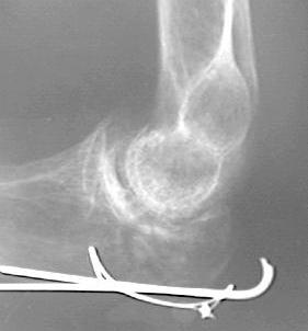
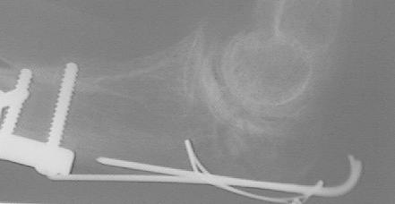
On June 1st 1998 he fell at work and sustain a fracture of the Left (ipsilateral) Olecranon Process. Immediate treatment consisted of open reduction and internal fixation of the olecranon fracture using standard tension band wiring technique. A bone biopsy was taken at the same time and showed features typical of Gorham's Disease. Ancillary treatment consisting of electrical stimulation of the fracture site and pamidronate infusion (60mg over 4 hours once weekly for 6 weeks) was begun once the diagnosis was confirmed.
 |
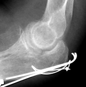 |
| 1 June 1998 Fracture of L olecranon through bone affected by Gorhams Disease | 9 June 1998 8 days post ORIF the fracture is compressed and only just visible |
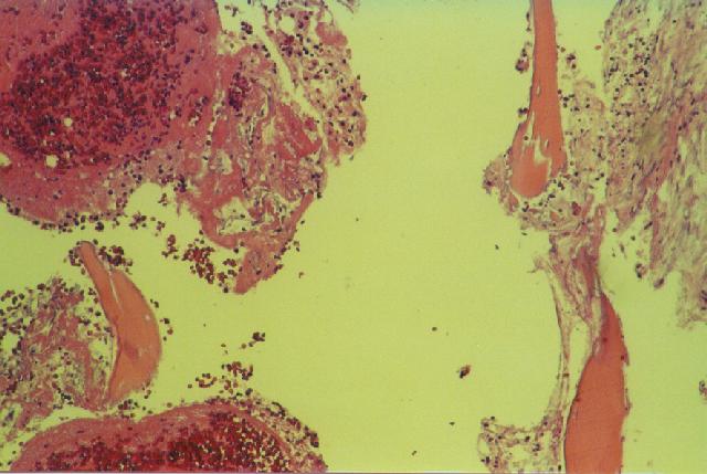 |
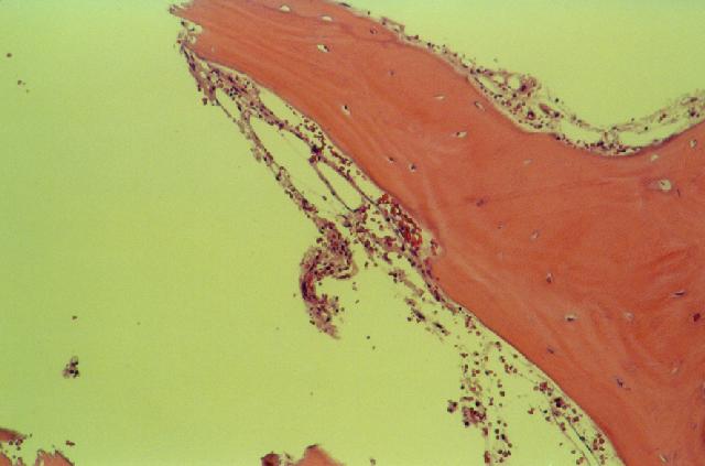
Biopsy from L Olecranon
Fracture June 1998 Haemorrhage and blood clot consistent with recent fracture. Background fibrillary marrow fibrosis and vascular ectasia in keeping with Gorham's disease |
 |
 |
 |
| July 22nd 1998 Radiolucency at the fracture line raises the possibility of activation of Gorham's Disease at the injury site | August 18th 1998 suggestion of bone filling in the fracture line may be due to differences in technique. The patient has returned to work against advice. Radiotherapy was initiated. |
 |
September 15th 1988 further evidence of
callus. Note on this view the radiolucencies that have developed around the proximal
screws of the old plate on the ulna. These lucencies have been noted from the June 1st
1998 xray. Follow-up to November 1998, 5 months post injury continues to indicate the fracture has healed. |
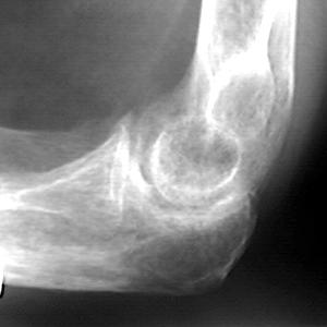 Lateral L elbow 16/Oct/1995